Tag Archives: biology

The Hypothalamus and Pituitary Gland, with Animation.

This video is available for licensing on our website. Click HERE!

The hypothalamus and the pituitary gland are at the center of endocrine function. The hypothalamus is part of the brain, while the pituitary, also called hypophysis (hy-POFF-ih-sis), is an endocrine gland. The hypothalamus links the nervous system to the endocrine system via the pituitary gland. The two structures are located at the base of the brain and are connected by a thin stalk.

The hypothalamus produces several hormones, known as neurohormones, which control the secretion of other hormones by the pituitary. Pituitary hormones, in turn, control the production of yet other hormones by other endocrine glands.

The pituitary has two distinct lobes:

The anterior pituitary, also called adenohypophysis (AD-eh-no-hy-POFF-ih-sis), communicates with the hypothalamus via a network of blood vessels known as the hypophyseal portal system. Several neurohormones produced by the hypothalamus are secreted into the portal system to reach the anterior pituitary, where they stimulate or inhibit production of pituitary hormones. Major hormones include:

  • Gonadotropin-releasing hormone, GnRH, a hypothalamic hormone, stimulates the anterior pituitary to producefollicle-stimulating hormone, FSH, and luteinizing hormone, FSH and LH, in turn, control the activities of the gonads – the ovaries and testes.
  • Corticotropin-releasing hormone, CRH,promotes the secretion of adrenocorticotropic hormone, ACTH, which in turn stimulates production of cortisol by the adrenal gland.
  • Thyrotropin-releasing hormone, TRH, promotes the release of thyroid-stimulating hormone, TSH, and prolactin. TSH, in turn, induces the thyroid gland to produce thyroid hormones. Prolactin stimulates the mammary glands to produce milk.
  • Prolactin-inhibiting hormone, PIH, inhibits production of prolactin.
  • Growth hormone–releasing hormone, GHRH, promotes production of growth hormone, or somatotropin, which has widespread effects on the growth of various tissues in the body.
  • Growth hormone–inhibiting hormone, GHIH, or somatostatin, inhibits production of growth hormone.

The posterior pituitary, also called neurohypophysis, communicates with the hypothalamus via a bundle of nerve fibers. These are essentially hypothalamic neurons with cell bodies located in the hypothalamus while their axons EXTENDED to posterior pituitary. These neurons produce hormones, transport them down the stalk, and store them at the nerve terminals within the posterior pituitary, where they wait for a nerve signal to trigger their release. Two hormones have been identified so far:

– Vasopressin, also known as antidiuretic hormone, ADH, acts on the kidneys to retain water.

– Oxytocin causes the uterus to contract during childbirth and stimulates contractions of the milk ducts in lactating women.

Email this to someoneShare on FacebookTweet about this on TwitterShare on Google+Share on LinkedIn

Protein Synthesis – Translation

Below is a narrated animation of prokaryotic translation. Click here to license this video on Alila Medical Media website.

Translation is the process of making polypeptide (protein) from the messenger RNA (mRNA).

The translation process involves the following components:
– mRNA or messenger RNA containing the genetic information to be translated.
– tRNA or transfer RNA bringing in the amino acids – the building blocks of the protein.
– Ribosome – the machine that performs the translation. The ribosome has two subunits: small and large.
– Several initiation factors (IFs), elongation factors (EFs) and release factors (RFs). These factors assist with initiation, elongation and termination of the process, respectively.

Steps of the translation process

Initiation: The small ribosomal subunit binds to the initiator tRNA carrying the initiator amino acid methionine (fMet). In eukaryotes, this complex then attaches to the cap structure at the 5’ end of an mRNA and scans for the start codon AUG. The process is mediated by several initiation factors. This is cap-dependent initiation. In some cases, the initiation complex binds to an internal ribosome entry sites (IRES) on the mRNA  – this is cap-independent initiation. The rest of the events remain the same. In prokaryotes, the initiation complex recognizes and binds to a  a purine-rich region – the Shine Dalgarno sequence –  upstream of the AUG initiation codon. 

At the start codon, the large ribosomal subunit joins the complex and all initiation factors are released. The ribosome has three sites: the A-site is the entry site for new tRNA charged with amino-acid or aminoacyl-tRNA; the P-site is occupied by peptidyl-tRNA – the tRNA that carries the growing polypeptide chain; the E-site is the exit site for the tRNA after it’s done delivering the amino acid. The initiator tRNA is positioned in the P-site.

Protein synthesis initiation (eukaryote)
Fig. 1: Translation initiation (eukaryotic, cap-dependent). Click on image to see it on Alila Medical Media website where the image is also available for licensing (together with other related images and videos).


Elongation: A new tRNA carrying an amino acid enters the A-site of the ribosome. On the ribosome, the anticodon of the incoming tRNA is matched against the mRNA codon positioned in the A-site. During this proof-reading, tRNA with incorrect anticodons are rejected and replaced by new tRNA that are again checked. When the right aminoacyl-tRNA enters the A-site, a peptide bond is made between the two now-adjacent amino-acids. As the peptide bond is formed, the tRNA in the P-site releases the amino-acids onto the tRNA in the A-site and becomes empty. At the same time, the ribosome moves one triplet forward on the mRNA. As a result, the empty tRNA is now in the E-site and the peptidyl tRNA moves to the P-site. The A-site is now unoccupied and is ready to accept a new tRNA. The cycle is repeated until the ribosome reaches a stop codon.
Protein synthesis elongation

Fig. 2: Translation elongation. Click on image to see it on Alila Medical Media website where the image is also available for licensing (together with other related images and videos).


Termination: Termination happens when one of the three stop codons is positioned in the A-site. No tRNA can fit in the A-site at that point as there are no tRNA that match that sequence. Instead, these codons are recognized by a protein, a release factor. Binding of the release factor catalyzes the cleavage of the bond between the polypeptide and the tRNA. The polypeptide is released from the ribosome. The ribosome is disassociated into subunits and is ready for a new round of translation. The newly made polypeptide usually requires additional modifications and folding before it can become an active protein.
Protein synthesis termination

Fig. 3: Translation termination. Click on image to see it on Alila Medical Media website where the image is also available for licensing (together with other related images and videos).

Email this to someoneShare on FacebookTweet about this on TwitterShare on Google+Share on LinkedIn

Molecular, Cell biology & Genetics Gallery

Download diagrams of normal human genome and chromosome changes in genetic diseases, illustrations of stem cells and cellular differentiation, gene expression and signaling, intracellular organelles, molecular techniques and more.

Please note: Free downloads are intended to facilitate healthcare education for people in need in low income countries and can be used for educational purposes only. If you can afford it or if you plan to use the images for commercial purposes, please consider buying instead. You can find a larger image collection at higher resolutions for sale at affordable prices on Alila Medical Media website.

To download: right click on full size image – choose “Save image as” and save it into your computer. By downloading from this website you acknowledge that you agree to our Conditions of Use.
To purchase larger sizes of the image: (left) click on full size image.

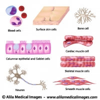
Human cell collection
Human cell collection, diagrams
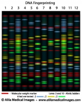
DNA fingerprinting, diagram.
DNA fingerprinting, in this example 6 loci are tested, molecular weight marker and allelic ladders added.
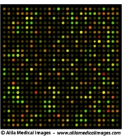
DNA microarrays chip, medical drawing.
Typical DNA chip results, technique used in (for example) cancer research to study differential expression of genes, illustration unlabeled.
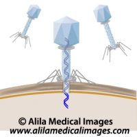
Phage infecting a bacterium, unlabeled diagram.
Phage injecting DNA into a bacterium, unlabeled drawing.
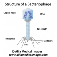
Structure of a bacteriophage, labeled diagram.
Structure of a phage, a virus that infects bacteria.
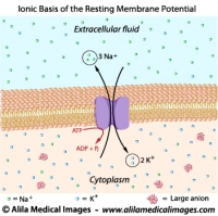
Ionic basis of resting membrane potential, labeled diagram.
Resting membrane potential is maintained by sodium potassium pump.
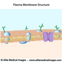
Structure of plasma membrane, unlabeled diagram.
Structure of cell membrane with lipid bilayer, cholesterol, transmembrane proteins and pores.
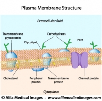
Structure of plasma membrane, labeled diagram.
Structure of cell membrane with lipid bilayer, cholesterol, transmembrane proteins and pores.
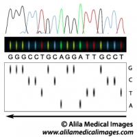
DNA sequencing principle, illustration.
DNA sequencing gel and interpretation of DNA sequence.
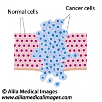
Cancer cells in a growing tumor, labeled diagram.
Cancer cells grow and break through the basement membrane, labeled drawing.
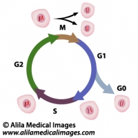
Cell cycle diagram.
Cell cycle diagram.
Phases of Cell cycle: mitosis, gap 1 (G1), gap 2 (G2), and S - DNA synthesis, G0 = quiescence.
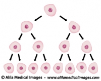
Cell division illustration.
Exponential growth of cells in a tissue, doubling of numbers of cells, unlabeled.
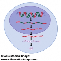
Gene expression, unlabeled diagram.
Gene expression process in eukaryotes, from transcription to protein folding.
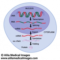
Gene expression, labeled diagram.
Gene expression process in eukaryotes, from transcription to protein folding.
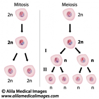
Mitosis versus meiosis, diagram.
comparing the two different types of cell division.
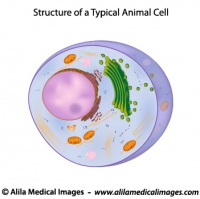
A typical cell, unlabeled diagram.
Structure of a typical eukaryotic cell, unlabeled drawing.
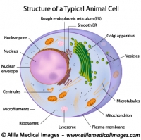
A typical cell, labeled diagram.
Structure of a typical eukaryotic cell, labeled drawing.
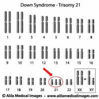
Down syndrome karyotype diagram.
Down syndrome karyotype, extra chromosome 21.
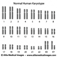
Normal human karyotype diagram.
Normal human diploid set of chromosomes.
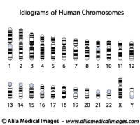
Human chromosome idiograms, diagrams
Accurate human chromosome banding patterns.
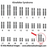
Klinefelter syndrome genome diagram.
Klinefelter syndrome genome has an extra X chromosome, XXY.
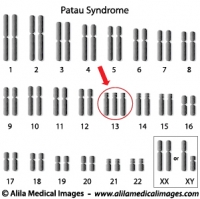
Patau syndrome, trisomy 13 diagram.
Patau syndrome genome has an extra chromosome 13.
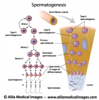
Spermatogenesis process, labeled diagram.
Process of sperm production and meiosis in seminiferous tubule, labeled diagram.
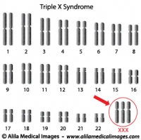
Triple X syndrome genome diagram.
Trisomy X syndrome genome has an extra X chromosome, female XXX.
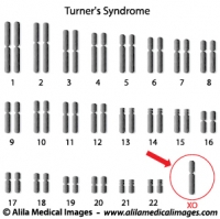
Turner's syndrome genome diagram.
Monosomy X, Turner's syndrome genotype is XO, one X chromosome is missing, female phenotype.
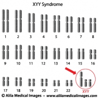
Super Male Syndrome genome diagram.
XYY, Super Male Syndrome genome has an extra Y chromosome.


Email this to someoneShare on FacebookTweet about this on TwitterShare on Google+Share on LinkedIn