Tag Archives: diseases

Piriformis syndrome


Piriformis syndrome (PS) is a neuromuscular condition where the piriformis muscle – one of the deep gluteal muscles – presses on and compresses the sciatic nerve causing pain, tingling and numbness in the buttock area, and down the path of sciatic nerve to the thigh and leg. Sciatic nerve runs under the piriformis muscle (Fig. 1) and may be irritated when the muscle is too tight or shortened due to spasms. Piriformis syndrome is to be differentiated from sciatica which shows similar symptoms but has different causes.

Piriformis syndrome

Fig. 1 : Piriformis syndrome. Posterior view of the pelvis showing location of piriformis muscle and sciatic nerve. Click on image to see a larger version on Alila Medical Media website where the image is also available for licensing.


Diagnosis is difficult as it produces similar symptoms as sciatica and is commonly done by exclusion of sciatica caused by compression of sciatic nerve roots by a herniated disc.

Lumbar spine disc herniation.
Fig. 2 : Sciatica caused by compression of spinal nerve roots by a herniated disc. Lateral view of the lumbar spine. Click on image to see a larger version on Alila Medical Media website where the image is also available for licensing.  


Causes and Risk factors

–  Anatomical abnormality of the nerve/muscle relation. Some people are more likely to get PS than others.

– Tightness or spasm of piriformis muscle due to overuse injury. This commonly happens in sport activities that put pressure on the piriformis muscle such as bicycling, running without proper stretching, or any activity that involves repeated movements of the leg performed in sitting position.

Treatment

– Conservative treatment includes stretching exercises, massage, avoidance of causative activities.

– Physical therapy that strengthens the gluteus maximus, gluteus medius, and biceps femoris is usually recommended to reduce strain on the piriformis muscle.

– Relief of symptoms may be achieved with anti-inflammatory drugs or muscle relaxants.


Email this to someoneShare on FacebookTweet about this on TwitterShare on Google+Share on LinkedIn

Femoral acetabular impingement (FAI)

What is femoral acetabular impingement (FAI)?

Femoral acetabular impingement (FAI) is a condition of hip joint where the bones are abnormally shaped, they pinch each other on the covering cartilages when the joint is in motion and cause damages.

The hip joint is a ball-and-socket joint  (Fig. 1). The femoral head (the ball) fits into the acetabulum (the socket). The femoral head is covered with articular cartilage, the acetabulum has a ring of cartilage around its rim called the labrum. In FAI, there are abnormal bone growths (spurs) on the ball or the socket or both (Fig. 2). The ball can no longer move smoothly inside the socket. They rub onto each other and pinch on the covering cartilages causing damages.

Hip joint structure, labeled.

Fig. 1 : Anatomy of the hip joint. Click on image to see a larger version on Alila Medical Media website where the image is also available for licensing.


Types of FAI

Pincer – Bone spurs on the acetabulum, commonly on the upper edge (Fig. 2 ). This situation is also described as over-coverage of the socket over the ball.

Cam – Bones spurs on the femoral head and neck making the neck less prominent and the head not completely round.

Combined – both cam and pincer are present, this is a very common situation.
Femoroacetabular impingement
Fig. 2 : Types of FAI. Bones spurs are colored in red. Click on image to see a larger version on Alila Medical Media website where the image is also available for licensing.


What damages can it make?

Impingement results in cartilage breakdown on the femoral head and labral tears on the rim of the acetabulum. FAI is also the cause of premature hip osteoarthritis in young adults.

Causes and risk factors

The bone spurs are the result of abnormal bone growth during childhood development. The reasons why this happens are unclear.

FAI is more common in young athletes, dancers who practice a larger range of motions of the hip, and in active individuals.


Symptoms

Some of the symptoms may include:

– Pain at the groin area or inner hip is more common although the pain may be felt at the side of the hip.

– Pain after sitting for a long period of time.

– Stabbing pain when sitting down or standing up.

Treatment

Treatments range from lifestyle changes, physical therapies to surgeries.

Lifestyle changes usually involve being less active. Physical therapy helps to increase hip strength but stretching should be avoided.

Arthroscopic surgeries are commonly performed to remove damaged tissues, repair the labrum and stimulate cartilage growth by microfractures. Removal of abnormal bony structures are also recommended to prevent future damages to the joint.

                                                                                                           >  See all Orthopedic topics

Email this to someoneShare on FacebookTweet about this on TwitterShare on Google+Share on LinkedIn

Benign Prostatic Hyperplasia (BPH)

Below is a narrated animation about BPH and treatments. Click here to license this video on Alila Medical Media website.

Benign prostatic hyperplasia  (BPH), also called benign prostatic hypertrophy or enlarged prostate,  is a condition in which the size of the prostate gland is increased. It is considered “benign” because it’s NOT a cancer, and it does not increase the risk of cancer. However, when becomes sufficiently large, the prostate tissue may compress the urethra and block the urine flow causing a number of urination problems and urinary tract infection. BPH is very common in aging men: about 50% of men have some degree of BPH by the age of 60.

Anatomy

The prostate is a walnut-size exocrine gland of the male reproductive system. It is located just below the urinary bladder where it wraps around the first part of the urethra – prostatic urethra (see Fig. 1 and 2).
Male reproductive system median section
Fig. 1: Male reproductive and urinary organs, mid-sagittal view. Click on image to see a larger version on Alila Medical Media website where the image is also available for licensing. 


Prostate gland produces a milky fluid that is expelled into the urethra to mix with spermatozoa during ejaculation. The fluid serves as a lubricant and nutrition for the sperms.

Click here to see an animation of male reproductive system on Alila Medical Media website where the video is also available for licensing.

Click here to see an animation of male urinary system on Alila Medical Media website where the video is also available for licensing.

In BPH, the enlarged prostate presses on the prostatic urethra making it narrower. This affects normal flow of urine (Fig. 2).

Benign prostatic hyperplasia (BPH)
Fig. 2: Normal prostate (left) and enlarged prostate (right). Same sagittal view as in Fig. 1 with other organs removed to simplify. The urethra is squeezed narrow in BPH.  Click on image to see a larger version on Alila Medical Media website where the image is also available for licensing.  


Click here to see  an animation of prostate hypertrophy on Alila Medical Media website where the video is also available for licensing.

Causes

BPH is considered a normal part of male aging as a result of hormonal changes. The rate of cell proliferation induced by androgens (male hormones) somehow exceeds the rate of programmed cell death (apoptosis) in aging prostate tissue resulting in enlargement of the prostate.
Severity of BPH (development of symptomatic BPH), however, has been associated with lifestyle. The incidence of clinically significant BPH is notably higher in men who lead a modern lifestyle compared to those who live in rural traditional settings.
About half of men with histopathologic BPH demonstrate clinically significant symptoms.

Symptoms

Obstruction of urine flow makes urine voiding difficult and incomplete. This leads to common symptoms of BPH:
– frequent urination.
– urgency : need to void that can not be deferred.
– urinary hesitation: difficulty to initiate urine stream, weak and interrupted stream.
– straining to void: need to push to completely empty the bladder.
– residual urine: constant feeling of need to void.
– dribbling
Altogether the voiding dysfunction resulted from BPH is called lower urinary tract symptoms (LUTS) – a more recent term for prostatism.

Treatment

Patients with mild symptoms and who are not bothered by their symptoms are usually advised to follow a “watch and see” approach with regular check-up and lifestyle changes such as low-fat diet, reduced consumption of alcohol and caffeine, reduced fluid intake before bedtime, avoidance of certain products and medications such as diuretics,…
1. Medication
There are two main classes of medication:
– alpha-blockers: these drugs relax smooth muscle in the prostate and by doing so relieve blockage of urine flow.
– 5-alpha reductase inhibitors: these inhibit local production of the hormone that is responsible for prostate enlargement.

2. Minimal invasive treatment
These non-surgical therapies use heat to cause cell death (necrosis) in prostate tissue. The heat is delivered in small amount and to a specific location to minimize unwanted damage. Different procedures differ mainly in the type of energy used.
– Transurethral microwave thermotherapy (TUMT): use of microwave energy delivered through a probe inside a catheter inserted into the urethra.
– Transurethral needle ablation (TUNA) : use of radio frequency energy delivered through a transurethral device with needles.
– Photoselective vaporization of the prostate (PVP): use of laser to vaporize prostate tissue.
3. Surgery
Transurethral resection of the prostate (TURP) is a surgical procedure for removal of prostate tissue through the urethra. This procedure has been around for a long time and is still considered gold standard for treatment of severe BPH. Nowadays, it is usually performed when medications and less invasive methods fail.

                                                                                                                    See all Urology topics

Email this to someoneShare on FacebookTweet about this on TwitterShare on Google+Share on LinkedIn

Asthma (with Animation Video)

The videos on this page can be downloaded upon purchase of a license on Alila Medical Media website. Click here!


Asthma is a chronic respiratory condition where the airway is inflamed and narrowed causing breathlessness, wheezing, chest tightness and coughing. Symptoms come as recurrent episodes called asthmatic attacks more commonly during the night and early morning. Asthma is usually diagnosed in childhood and lasts for life.

Anatomy of asthma

Our lungs consist of millions of air tubes or airways (bronchi and smaller bronchioles) bringing air in and out of the body. Each tube ends with a cluster of air sacs (alveolus, plural alveoli) where the gas exchange process takes place. The airways have a layer of smooth muscle in their wall which enables them to constrict or dilate. In response to the body’s higher demand of air, such as during exercise, the airways dilate to increase air flow. In response to presence of pollutants in the air, they constrict to prevent the lungs from being polluted. In people suffering from asthma these airways are inflamed, narrowed and become more sensitive to certain substances. Asthmatic attack (or exacerbation) happens when the airways react to these substances. During the attack smooth muscle contracts squeezing the airways making them even narrower, mucus secretion increases further obstructs the airways.

Causes of asthma

Causes of asthma are complex and not fully understood but likely involve a combination of genetic and environmental factors. Family history is a known risk factor for asthma. There are at least over twenty genes associated with asthma of which many are involved in the immune system. Most people who have asthma also have allergies. Many environmental factors such as air pollution, chemicals, smoking, allergens have been associated with development of asthma or triggering of asthmatic attacks.

Triggers of asthmatic attack

Triggers are factors that initiate the attack, these can be very different from person to person. Common triggers include :
– allergens (pollen, animal fur, pet dander, sulfites in preserved food..)
– irritants (cigarette smoke, industrial chemicals, dust, household chemicals,..)
– some medication (aspirin, beta blockers,..).
– physical activity, exercise.

Treatments

There is no cure for asthma. The most effective way to manage symptoms is to identify the triggers of asthmatic attack and avoid them.
There are two main classes of medication:
– bronchodilators – substances that dilate bronchi and bronchioles – are used as short-term relief of symptoms.
– inflammation moderators such as corticosteroids are used as long-term treatment.
Asthma inhalers are used to deliver medication to the lungs.

Associated conditions

A number of conditions tend to occur more frequently in people with asthma:
– Allergies :  eczema and hay fever. These individuals are considered hyperallergic (high tendency to develop allergic reactions). The combination of these conditions is called atopy or atopic symdrome.
Gastroesophageal reflux disease (GERD): a condition in which stomach acid backs up and damages the mucosal lining of the esophagus. GERD may worsen asthma symptoms and medications for asthma often worsen GERD symptoms. Treating GERD usually improves asthma and must be included in asthma treatment plan.
Obstructive sleep apnea (OSA): asthmatic patients tend to develop OSA. The mechanism is not fully understood but it’s likely due to nasal obstruction. Click on the link to read more about OSA.
Sinusitis: inflammation of paranasal sinuses. Sinusitis commonly worsens asthma symptoms and makes treatment less effective.

                                                                                                        See all Respiratory topics

                                                                                                        See all Immunology topics
 

Email this to someoneShare on FacebookTweet about this on TwitterShare on Google+Share on LinkedIn

GERD and Heartburn (with video)

Gastroesophageal reflux disease

This video and other animations of the digestive system are available for licensing on Alila Medical Media website. Click here!


Gastroesophageal reflux disease (GERD) or gastric reflux disease is  a chronic condition where acid from the stomach flows up and damages the mucosa of the esophagus.
At the junction between the esophagus and the stomach is the lower esophageal sphincter (LES). The LES is a ring of muscle that is generally closed tight to prevent stomach acid from coming up. In normal digestion, the LES opens shortly to allow food bolus passing down to the stomach and closes back tight instantly. GERD occurs when the LES is abnormally relaxed and can not close properly (Fig. 1). Heartburn is a burning sensation in the chest associated with each regurgitation of gastric acid and is the most prominent symptom of GERD. 

Gastric reflux, labeled diagram.
Fig. 1: Abnormal relaxation of the lower esophageal sphincter as cause of GERD. Click on image to see a larger version on  Alila Medical Media website where the image is also available for licensing.


Hiatus hernia is believed to be another cause of GERD. Hiatus hernia or hiatal hernia is a condition where the top portion of the stomach is pulled up forming a herniation above  the diaphragm. This situation somehow compromises the esophagus – stomach barrier and facilitates acid reflux.

Hiatal Hernia
Fig. 2: Types of hiatal hernia. Click on image to see a larger version on  Alila Medical Media website where the image is also available for licensing.


Treatment

Treatment includes dieting and medication. Proton-pump inhibitors, which act to reduce gastric acid production, are drugs of choice. If these fail, a surgery may be recommended. In a procedure called Nissen fundoplication, the top portion of the stomach is wrapped around the lower part of esophagus and sewn into place. This way, the muscles in the wall of the stomach reinforce the closure of the esophagus. This surgical procedure is particularly recommended when hiatus hernia is present as this can be fixed at the same time. The procedure can be done with minimal invasive laparoscopic technique through small incisions with the aid of a camera.
Nissen Fundoplication Surgery
Fig. 3: Nissen fundoplication procedure.  Click on image to see a larger version on Alila Medical Media website where the image is also available for licensing.


                                                                                                                 > See all Digestive topics
Email this to someoneShare on FacebookTweet about this on TwitterShare on Google+Share on LinkedIn

Common shoulder injuries and surgical repair (part 2)

Rotator cuff injuries


Fig. 1 shows a group of four muscles that cover the shoulder joint. These muscles originate on the scapula and insert on the humerus: the supraspinatus,  infraspinatus, subscapularis and teres minor. The tendons of theses muscles form the rotator cuff (tendons connect muscles to bones). The most common injury to the rotator cuff is the impingement of one or more of these tendons. This may happen as a result of  a trauma or sport related injury but more commonly as a result of aging. The tendons may rub against the acromion (a bony extension of the scapula that hangs over the cuff) every time the person raises an arm and become irritated, inflamed and ultimately torn.

Rotator cuff muscles
Fig. 1: Rotator cuff muscles of the right shoulder. Click on image to see a larger version on Alila Medical Media website where the image is also available for licensing.


Below is a narrated animation of arthroscopic rotator cuff repair.  Click here to license this video and/or other orthopedic videos on Alila Medical Media website.

Impingement usually develops over a period of time. Treatment includes rest, shoulder exercise, physical therapy and surgery. In most cases surgical treatment is done through an arthroscope but open surgery may be needed for larger tears. During surgery the damaged tissue is removed, source of irritation (commonly bone spurs on the acromion) is identified and removed. If there is no tear, the treatment may stop here and the surgical procedure is called shoulder debridement. In case of tear sutures will be used to tight the tendon back down to the bone (Fig.2).

Rotator cuff repair diagram.
Fig 2. Rotator cuff injury (1) and repair: small holes are drilled (2) into the bone of the humerus to hold small suture anchors with threads (3). The threads are attached to the tendon (4) and pulled tightly to hold the tendon to the bone (5). Click on image to see a larger version on Alila Medical Media website where the image is also available for licensing.

Separated shoulder

Separated shoulder is a condition affecting the “second” joint of the shoulder : the AC joint (acromioclavicular joint) between the acromion (an extension of the scapula)  and the clavicle. This condition is commonly due to a direct blow to the shoulder as in a fall or sport injury. Fig. 3 shows the ligaments involved in stabilization of AC joint, injury to any of these ligament results in separated shoulder. Injuries are graded according to the extend of tears and number of ligaments involved. Grade I injury (partial tear in one of the ligament) may be treated with simple rest and ice, small tears heal themselves over time. Grade III injury where the clavicle is completely detached from the scapula requires surgery where a screw will be inserted to fix the clavicle to the coracoid process of the scapula.

Separated shoulder, labeled diagram.
Fig.3 : Shoulder separation grading. Click on image to see a larger version on Alila Medical Media website where the image is also available for licensing.

Frozen shoulder (adhesive capsulitis)

The shoulder, like all synovial joint, has a capsule around it. The capsule encloses the two end surfaces of the bones involved in the joint and a joint cavity containing a lubricant called synovial fluid. In people with frozen shoulder condition this capsule is thicken and inflamed (Fig. 4) causing pain when they try to move an arm. The pain increases with time and the range of motion decreases, the shoulder becomes stiff or “frozen”.
Adhesive capsulitis of shoulder diagram.
Fig. 4: Frozen shoulder. Click on image to see a larger version on Alila Medical Media website where the image is also available for licensing.


The causes of frozen shoulder are not fully established. People with diabetes and some other diseases show increased risk for frozen shoulder. It can also be resulted from a long-term immobilization of the shoulder (for example after a shoulder surgery). Treatments include pain management and physical therapy although in some cases surgery may be necessary. A procedure called arthroscopic capsular release  is usually performed to cut through the tight area of the capsule.

                                                                                                              >  See all Orthopedic topics
Email this to someoneShare on FacebookTweet about this on TwitterShare on Google+Share on LinkedIn

Common shoulder injuries and surgical repair (part 1)

Anatomy


There are three bones in the shoulder: the humerus (the bone of the upper arm), the scapula (shoulder blade) and the clavicle (collarbone). Articulations between these bones make up the shoulder joints. The main joint, commonly referred to as “the shoulder joint”, is the joint between the head of the humerus and glenoid cavity of the scapula and is called the humeroscapular or glenohumeral joint. The second joint of the shoulder is formed by the articulation between the clavicle and the acromion (extension of the scapula that forms the top of the shoulder) and is called acromioclavicular joint or AC joint. The two joints are stabilized by associated muscles and ligaments.

Shoulder anatomy
Fig.1: Main components of the shoulder joint. Click on image to see a larger version on  Alila Medical Media website where the image is also available for licensing.


Shoulder dislocation

Shoulder dislocation occurs when the humeral head slips out of the pocket  made by glenoid cavity of the scapula (Fig. 2). This usually happens as a result of trauma (fall, sport injury,..). Dislocation can be anterior where the humerus slips to the front or posterior where it dislocates behind the normal position. Anterior dislocation is more common.

Shoulder dislocation
Fig.2 : Types of shoulder dislocation. Click on image to see a larger version on Alila Medical Media website where the image is also available for licensing.


Below is a narrated animation about shoulder dislocation, bankart lesion and repair. Click here to license this video and/or other orthopaedic videos on Alila Medical Media website.

Bankart lesion and shoulder instability

The glenoid cavity has a ring of fibrocartilage tissue called the labrum around it. The labrum makes the cavity deeper and helps to keep the humeral head in place (Fig.3). During anterior shoulder dislocation, the head of the humerus may be pressed against and damages the anterior portion of the labrum. This type of labral tear is called bankart. Damaged labrum makes it easier for the humeral head to slip out of place again. This vicious cycle leads to repeated shoulder dislocation and severely damaged labrum. The condition is called shoulder instability as it feels like slipping out anytime. Treatment includes physical therapy and, in some cases, surgery for bankart repair (see below).

Click here to see an animation of bankart lesion and arthroscopic repair  on Alila Medical Media website where the video is also available for licensing.

SLAP and bankart lesions
Fig. 3: Anatomy of the shoulder joint with the humerus slightly abducted to show the glenoid cavity and labrum. Types of labral lesions are shown on the right. Click on image to see a larger version on Alila Medical Media website where the image is also available for licensing.


Bankart repair

During surgery the damaged cartilage is removed, area is cleaned, small holes are then drilled into the bone of glenoid fossa to hold small suture anchors with threads. The threads are attached to the labrum and pulled tightly to hold the labrum to the glenoid (Fig.4). Over time, the labrum will reattach to the glenoid naturally. Physical therapy will be needed to regain the shoulder range of motions and strength.

Shoulder stabilization surgery
Fig.4: Steps of bankart repair surgery. See text for details. Click on image to see a larger version on Alila Medical Media website where the image is also available for licensing.


Bankart repair can be done with arthroscopic or open surgery. While arthroscopy is minimal invasive, open surgery maybe recommended for larger tears. In arthroscopic surgery only two or three small incisions are made, an arthroscope is inserted through one of the incision. Arthroscope is an instrument equipped with light and camera which transmits image of the joint to a computer screen. Other small tools are inserted  to carry out the repair (Fig.5).

Click here to see an animation of arthroscopic bankart repair.
Shoulder arthroscopy
Fig. 5 : Arthroscopic surgery for bankart repair. Click on image to see a larger version on Alila Medical Media website where the image is also available for licensing.


                                                                                                              >  See all Orthopedic topics
Email this to someoneShare on FacebookTweet about this on TwitterShare on Google+Share on LinkedIn

Male Reproductive System Gallery

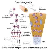
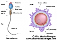
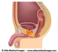
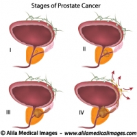
Download diagrams of male organs anatomy and diseases, formation and structure of sperms.

Please note: Free downloads are intended to facilitate healthcare education for people in need in low income countries and can be used for educational purposes only. If you can afford it or if you plan to use the images for commercial purposes, please consider buying instead. You can find a larger image collection at higher resolution for sale at affordable prices on Alila Medical Media website.

To download: right click on full size image – choose “Save image as” and save it into your computer. By downloading from this website you acknowledge that you agree to our Conditions of Use.
To purchase larger sizes of the image: (left) click on full size image.

Spermatogenesis process, labeled diagram.
Process of sperm production and meiosis in seminiferous tubule, labeled diagram.
Ovum and spermatozoon structure, labeled diagram.
Egg and sperm cells anatomy, labeled drawing.
Male reproductive organs sagittal, unlabeled diagram.
Illustration of male reproductive system in median section
Male reproductive organs sagittal, labeled diagram.
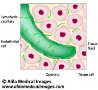
Illustration of male reproductive system in median section
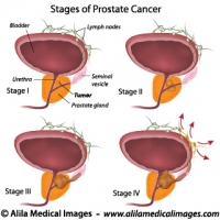
Prostate cancer staging, unlabeled diagram.
Illustration of staging of prostate gland cancer, from small tumor size to metastasis.
Prostate cancer staging, labeled diagram.
Illustration of staging of prostate gland cancer, from small tumor size to metastasis.


Email this to someoneShare on FacebookTweet about this on TwitterShare on Google+Share on LinkedIn

Lymphatic System Gallery

Download illustrations of major groups of lymph nodes, lymphatic organs and vessels, their anatomy, function, disorders and involvement in cancer metastasis.

Please note: Free downloads are intended to facilitate healthcare education for people in need in low income countries and can be used for educational purposes only. If you can afford it or if you plan to use the images for commercial purposes, please consider buying instead. You can find a larger image collection at higher resolution for sale at affordable prices on Alila Medical Media website.

To download: right click on full size image – choose “Save image as” and save it into your computer. By downloading from this website you acknowledge that you agree to our Conditions of Use.
To purchase larger sizes of the image: (left) click on full size image.

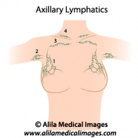
Axillary lymph nodes, medical drawing.
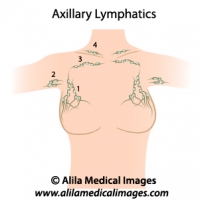
Axillary lymph nodes diagram, simple version. 1-3 = different groups of axillary nodes, 4= supraclavicular lymph nodes. Unlabeled diagram.
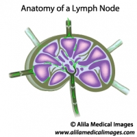
Lymph node structure, unlabeled diagram.
Diagram illustrating structure of a lymph node with lymph flow, unlabeled.
Lymph node structure, labeled diagram.
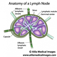
Diagram illustrating structure of a lymph node with lymph flow, labeled.
Lymph nodes of head and neck, medical drawing.
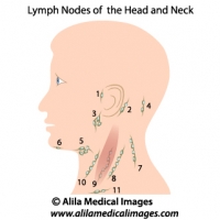
Lymph nodes of the head and neck, unlabeled. 1= preauricular, 2= posterior auricular, 3= parotid, 4= occipital, 5= submandibular, 6= submental, 7-10 = cervical, 11= supraclavicular.
Lymphatic fluid drainage, labeled diagram.
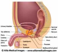
Uptake of tissue fluid by lymphatic capillary, labeled drawing.
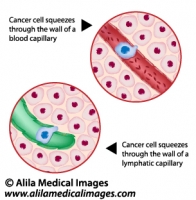
Metastatic cancer, medical drawing.
Cancer cell squeezes through the endothelium of blood and lymph vessel, illustration unlabeled.
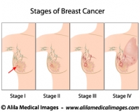
Breast cancer staging, diagram.
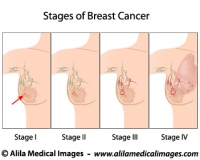
Stages of Breast Cancer, from small tumor to metastasis, medical illustration, unlabeled.
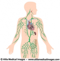
Lymphatic system, unlabeled diagram.
Human lymphatic system with all organs, major trunks, ducts and groups of lymph nodes illustrated, unlabeled illustration.
Lymphatic system, labeled diagram.
Human lymphatic system with all organs, major trunks, ducts and groups of lymph nodes illustrated, labeled illustration.


Email this to someoneShare on FacebookTweet about this on TwitterShare on Google+Share on LinkedIn

Female Reproductive System Gallery

Download cliparts illustrating anatomy of reproduction organs, oogenesis and sexual cycle, hormonal control, women’s diseases and conditions.

Please note: Free downloads are intended to facilitate healthcare education for people in need in low income countries and can be used for educational purposes only. If you can afford it or if you plan to use the images for commercial purposes, please consider buying instead. You can find a larger image collection at higher resolution for sale at affordable prices on Alila Medical Media website.

To download: right click on full size image – choose “Save image as” and save it into your computer. By downloading from this website you acknowledge that you agree to our Conditions of Use.
To purchase larger sizes of the image: (left) click on full size image.

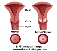
Cervical cancer, labeled diagram.
Diagram showing cancer of the cervix, labeled.
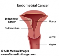
Uterine cancer, labeled diagram.
Illustration of tumor of the endometrium of female uterus, labeled.
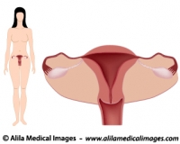
Women's system simple version, unlabeled.
Diagram showing female body with anatomy of reproductive system, anterior view, simple unlabeled version.
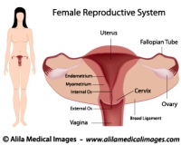
Women's system simple version, labeled.
Diagram showing female body with anatomy of reproductive system, anterior view, simple labeled version.
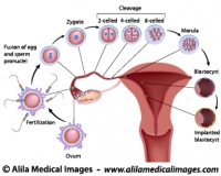
From fertilization to implantation, labeled diagram.
Diagram showing early human embryo development events, from fertilization to implantation of blastocyst, labeled.
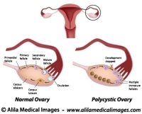
Polycystic and normal ovary, labeled diagram.
Normal ovarian cycle and Polycystic ovary syndrome illustration, labeled.
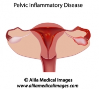
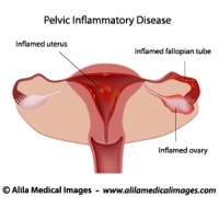
Pelvic inflammatory disease (PID)
Pelvic inflammatory disease (PID) diagram, unlabeled
Pelvic inflammatory disease (PID) diagram
Pelvic inflammatory disease (PID) diagram, labeled
Axillary lymph nodes, medical drawing.
Axillary lymph nodes diagram, simple version. 1-3 = different groups of axillary nodes, 4= supraclavicular lymph nodes. Unlabeled diagram.
Breast cancer staging, diagram.
Stages of Breast Cancer, from small tumor to metastasis, medical illustration, unlabeled.
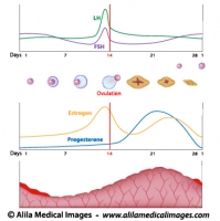
Menstrual cycle and ovulation, labeled diagram.
Normal menstrual cycle and the hormonal control of ovulation, labeled illustration.
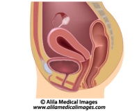
Women's reproductive system, unlabeled diargam.
Diagram showing organs of female reproductive tract in median section.
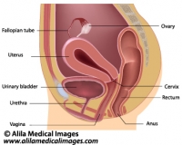
Women's reproductive system, labeled diargam.
Diagram showing organs of female reproductive tract in median section.
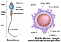
Ovum and spermatozoon structure, labeled diagram.
Egg and sperm cells anatomy, labeled drawing.


Email this to someoneShare on FacebookTweet about this on TwitterShare on Google+Share on LinkedIn