Tag Archives: drawing

Common shoulder injuries and surgical repair (part 2)

Rotator cuff injuries


Fig. 1 shows a group of four muscles that cover the shoulder joint. These muscles originate on the scapula and insert on the humerus: the supraspinatus,  infraspinatus, subscapularis and teres minor. The tendons of theses muscles form the rotator cuff (tendons connect muscles to bones). The most common injury to the rotator cuff is the impingement of one or more of these tendons. This may happen as a result of  a trauma or sport related injury but more commonly as a result of aging. The tendons may rub against the acromion (a bony extension of the scapula that hangs over the cuff) every time the person raises an arm and become irritated, inflamed and ultimately torn.

Rotator cuff muscles
Fig. 1: Rotator cuff muscles of the right shoulder. Click on image to see a larger version on Alila Medical Media website where the image is also available for licensing.


Below is a narrated animation of arthroscopic rotator cuff repair.  Click here to license this video and/or other orthopedic videos on Alila Medical Media website.

Impingement usually develops over a period of time. Treatment includes rest, shoulder exercise, physical therapy and surgery. In most cases surgical treatment is done through an arthroscope but open surgery may be needed for larger tears. During surgery the damaged tissue is removed, source of irritation (commonly bone spurs on the acromion) is identified and removed. If there is no tear, the treatment may stop here and the surgical procedure is called shoulder debridement. In case of tear sutures will be used to tight the tendon back down to the bone (Fig.2).

Rotator cuff repair diagram.
Fig 2. Rotator cuff injury (1) and repair: small holes are drilled (2) into the bone of the humerus to hold small suture anchors with threads (3). The threads are attached to the tendon (4) and pulled tightly to hold the tendon to the bone (5). Click on image to see a larger version on Alila Medical Media website where the image is also available for licensing.

Separated shoulder

Separated shoulder is a condition affecting the “second” joint of the shoulder : the AC joint (acromioclavicular joint) between the acromion (an extension of the scapula)  and the clavicle. This condition is commonly due to a direct blow to the shoulder as in a fall or sport injury. Fig. 3 shows the ligaments involved in stabilization of AC joint, injury to any of these ligament results in separated shoulder. Injuries are graded according to the extend of tears and number of ligaments involved. Grade I injury (partial tear in one of the ligament) may be treated with simple rest and ice, small tears heal themselves over time. Grade III injury where the clavicle is completely detached from the scapula requires surgery where a screw will be inserted to fix the clavicle to the coracoid process of the scapula.

Separated shoulder, labeled diagram.
Fig.3 : Shoulder separation grading. Click on image to see a larger version on Alila Medical Media website where the image is also available for licensing.

Frozen shoulder (adhesive capsulitis)

The shoulder, like all synovial joint, has a capsule around it. The capsule encloses the two end surfaces of the bones involved in the joint and a joint cavity containing a lubricant called synovial fluid. In people with frozen shoulder condition this capsule is thicken and inflamed (Fig. 4) causing pain when they try to move an arm. The pain increases with time and the range of motion decreases, the shoulder becomes stiff or “frozen”.
Adhesive capsulitis of shoulder diagram.
Fig. 4: Frozen shoulder. Click on image to see a larger version on Alila Medical Media website where the image is also available for licensing.


The causes of frozen shoulder are not fully established. People with diabetes and some other diseases show increased risk for frozen shoulder. It can also be resulted from a long-term immobilization of the shoulder (for example after a shoulder surgery). Treatments include pain management and physical therapy although in some cases surgery may be necessary. A procedure called arthroscopic capsular release  is usually performed to cut through the tight area of the capsule.

                                                                                                              >  See all Orthopedic topics

Email this to someoneShare on FacebookTweet about this on TwitterShare on Google+Share on LinkedIn

Male Reproductive System Gallery

Download diagrams of male organs anatomy and diseases, formation and structure of sperms.

Please note: Free downloads are intended to facilitate healthcare education for people in need in low income countries and can be used for educational purposes only. If you can afford it or if you plan to use the images for commercial purposes, please consider buying instead. You can find a larger image collection at higher resolution for sale at affordable prices on Alila Medical Media website.
To download: right click on full size image – choose “Save image as” and save it into your computer. By downloading from this website you acknowledge that you agree to our Conditions of Use.
To purchase larger sizes of the image: (left) click on full size image.
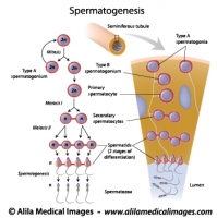
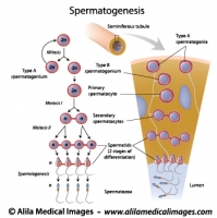
Spermatogenesis process, labeled diagram.
Process of sperm production and meiosis in seminiferous tubule, labeled diagram.
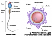
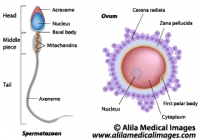
Ovum and spermatozoon structure, labeled diagram.
Egg and sperm cells anatomy, labeled drawing.
Male reproductive organs sagittal, unlabeled diagram.
Illustration of male reproductive system in median section
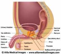
Male reproductive organs sagittal, labeled diagram.
Illustration of male reproductive system in median section
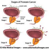
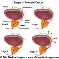
Prostate cancer staging, unlabeled diagram.
Illustration of staging of prostate gland cancer, from small tumor size to metastasis.
Prostate cancer staging, labeled diagram.
Illustration of staging of prostate gland cancer, from small tumor size to metastasis.


Email this to someoneShare on FacebookTweet about this on TwitterShare on Google+Share on LinkedIn

Lymphatic System Gallery

Download illustrations of major groups of lymph nodes, lymphatic organs and vessels, their anatomy, function, disorders and involvement in cancer metastasis.

Please note: Free downloads are intended to facilitate healthcare education for people in need in low income countries and can be used for educational purposes only. If you can afford it or if you plan to use the images for commercial purposes, please consider buying instead. You can find a larger image collection at higher resolution for sale at affordable prices on Alila Medical Media website.

To download: right click on full size image – choose “Save image as” and save it into your computer. By downloading from this website you acknowledge that you agree to our Conditions of Use.
To purchase larger sizes of the image: (left) click on full size image.

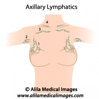
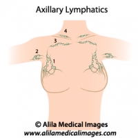
Axillary lymph nodes, medical drawing.
Axillary lymph nodes diagram, simple version. 1-3 = different groups of axillary nodes, 4= supraclavicular lymph nodes. Unlabeled diagram.
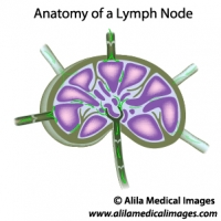
Lymph node structure, unlabeled diagram.
Diagram illustrating structure of a lymph node with lymph flow, unlabeled.
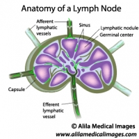
Lymph node structure, labeled diagram.
Diagram illustrating structure of a lymph node with lymph flow, labeled.
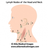
Lymph nodes of head and neck, medical drawing.
Lymph nodes of the head and neck, unlabeled. 1= preauricular, 2= posterior auricular, 3= parotid, 4= occipital, 5= submandibular, 6= submental, 7-10 = cervical, 11= supraclavicular.
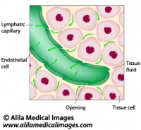
Lymphatic fluid drainage, labeled diagram.
Uptake of tissue fluid by lymphatic capillary, labeled drawing.
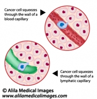
Metastatic cancer, medical drawing.
Cancer cell squeezes through the endothelium of blood and lymph vessel, illustration unlabeled.
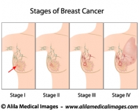
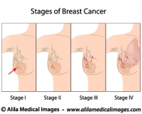
Breast cancer staging, diagram.
Stages of Breast Cancer, from small tumor to metastasis, medical illustration, unlabeled.
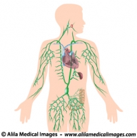
Lymphatic system, unlabeled diagram.
Human lymphatic system with all organs, major trunks, ducts and groups of lymph nodes illustrated, unlabeled illustration.
Lymphatic system, labeled diagram.
Human lymphatic system with all organs, major trunks, ducts and groups of lymph nodes illustrated, labeled illustration.


Email this to someoneShare on FacebookTweet about this on TwitterShare on Google+Share on LinkedIn

Female Reproductive System Gallery

Download cliparts illustrating anatomy of reproduction organs, oogenesis and sexual cycle, hormonal control, women’s diseases and conditions.

Please note: Free downloads are intended to facilitate healthcare education for people in need in low income countries and can be used for educational purposes only. If you can afford it or if you plan to use the images for commercial purposes, please consider buying instead. You can find a larger image collection at higher resolution for sale at affordable prices on Alila Medical Media website.

To download: right click on full size image – choose “Save image as” and save it into your computer. By downloading from this website you acknowledge that you agree to our Conditions of Use.
To purchase larger sizes of the image: (left) click on full size image.

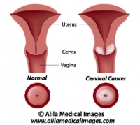
Cervical cancer, labeled diagram.
Diagram showing cancer of the cervix, labeled.
Uterine cancer, labeled diagram.
Illustration of tumor of the endometrium of female uterus, labeled.
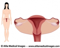
Women's system simple version, unlabeled.
Diagram showing female body with anatomy of reproductive system, anterior view, simple unlabeled version.
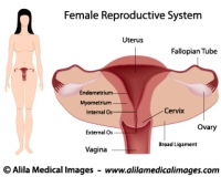
Women's system simple version, labeled.
Diagram showing female body with anatomy of reproductive system, anterior view, simple labeled version.
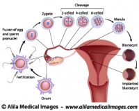
From fertilization to implantation, labeled diagram.
Diagram showing early human embryo development events, from fertilization to implantation of blastocyst, labeled.
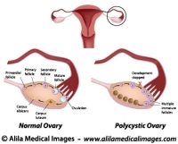
Polycystic and normal ovary, labeled diagram.
Normal ovarian cycle and Polycystic ovary syndrome illustration, labeled.
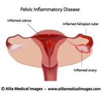
Pelvic inflammatory disease (PID)
Pelvic inflammatory disease (PID) diagram, unlabeled
Pelvic inflammatory disease (PID) diagram
Pelvic inflammatory disease (PID) diagram, labeled
Axillary lymph nodes, medical drawing.
Axillary lymph nodes diagram, simple version. 1-3 = different groups of axillary nodes, 4= supraclavicular lymph nodes. Unlabeled diagram.
Breast cancer staging, diagram.
Stages of Breast Cancer, from small tumor to metastasis, medical illustration, unlabeled.
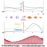
Menstrual cycle and ovulation, labeled diagram.
Normal menstrual cycle and the hormonal control of ovulation, labeled illustration.
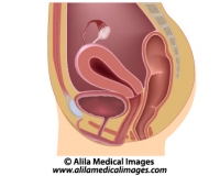
Women's reproductive system, unlabeled diargam.
Diagram showing organs of female reproductive tract in median section.
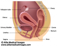
Women's reproductive system, labeled diargam.
Diagram showing organs of female reproductive tract in median section.
Ovum and spermatozoon structure, labeled diagram.
Egg and sperm cells anatomy, labeled drawing.


Email this to someoneShare on FacebookTweet about this on TwitterShare on Google+Share on LinkedIn

Oral Cavity Gallery

Download illustrations of permanent and deciduous dentition, tooth anatomy, periodontal diseases, teeth replacement therapy, mouth anatomy and diseases.

Please note: Free downloads are intended to facilitate healthcare education for people in need in low income countries and can be used for educational purposes only. If you can afford it or if you plan to use the images for commercial purposes, please consider buying instead. You can find a larger image collection at higher resolution for sale at affordable prices on Alila Medical Media website.

To download: right click on full size image – choose “Save image as” and save it into your computer. By downloading from this website you acknowledge that you agree to our Conditions of Use.
To purchase larger sizes of the image: (left) click on full size image.

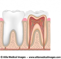
Tooth anatomy, unlabeled diagram.
Tooth anatomy from outside and in cross section, unlabeled drawing.
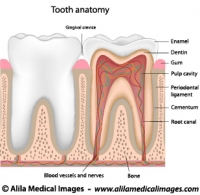
Tooth anatomy, labeled diagram.
Tooth anatomy from outside and in cross section, labeled drawing.
Dental checkup icon
Dental checkup icon
Magnifying glass on a molar tooth, illustration.
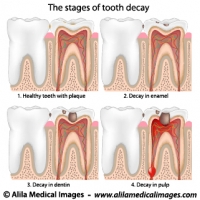
Plaque and tooth decay, medical drawing.
Tooth decay stages diagram.
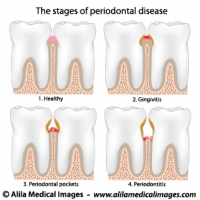
Periodontal Disease, labeled diagram.
Gum disease stages, from gingivitis to periodontitis.
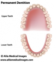
Adult dentition, medical drawing.
Permanent teeth, adult dentition, upper and lower jaws, unlabeled.
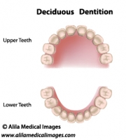
Deciduous dentition, medical drawing.
Deciduous dentition (baby or milk teeth), upper and lower jaws, medical illustration unlabeled..
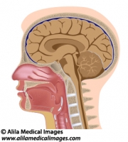
Head anatomy median section, medical drawing.
Median section of human head diagram with details of brain, sinuses, tonsils, oral and nasal cavity, pharynx and larynx, unlabeled.
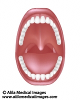
Mouth and throat anatomy, medical drawing.
Diagram of human oral cavity with denture, tonsils, unlabeled.


Email this to someoneShare on FacebookTweet about this on TwitterShare on Google+Share on LinkedIn

Urinary System Gallery

Download anatomical illustrations of kidneys, urine formation process, urinary tract diseases and conditions.

Please note: Free downloads are intended to facilitate healthcare education for people in need in low income countries and can be used for educational purposes only. If you can afford it or if you plan to use the images for commercial purposes, please consider buying instead. You can find a larger image collection at higher resolution for sale at affordable prices on Alila Medical Media website.

To download: right click on full size image – choose “Save image as” and save it into your computer. By downloading from this website you acknowledge that you agree to our Conditions of Use.

To purchase larger sizes of the image: (left) click on full size image.

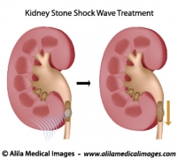
Treatment of kidney stones, medical drawing.
Ultrasound shock wave therapy for kidney stones, unlabeled.
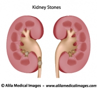
Kidney stones, medical drawing.
Diagram of healthy kidney anatomy and kidney with stones blocking urine passage, unlabeled.
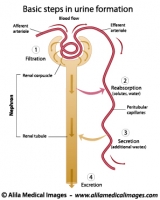
Formation of urine, labeled diagram.
Basic steps in urine formation in a nephron of kidney.
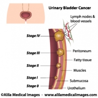
Urinary bladder cancer stages, medical drawing.
Staging of urinary bladder cancer diagram, unlabeled
Urinary bladder cancer stages, labeled.
Staging of urinary bladder cancer diagram, labeled
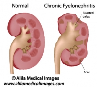
Chronic pyelonephritis, labeled diagram.
Chronic pyelonephritis kidney disease with tissue scarring and blunted calyx, medical illustration.
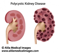
Polycystic kidney disease, medical drawing.
Diagram of enlarged kidney with multiple cysts compared to normal anatomy, unlabeled.
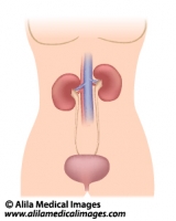
Urinary organs in female, medical illustration.
Female urinary system diagram showing kidneys, ureters and bladder, simple version unlabeled.
Prostate cancer staging, unlabeled diagram.
Illustration of staging of prostate gland cancer, from small tumor size to metastasis.
Prostate cancer staging, labeled diagram.
Illustration of staging of prostate gland cancer, from small tumor size to metastasis.


Email this to someoneShare on FacebookTweet about this on TwitterShare on Google+Share on LinkedIn

Eyes, Vision Gallery

Download diagrams of human eye anatomy, tear film and lacrimal gland, muscles of the orbit and associated nerves, defects and diseases, laser surgery treatment.

Please note: Free downloads are intended to facilitate healthcare education for people in need in low income countries and can be used for educational purposes only. If you can afford it or if you plan to use the images for commercial purposes, please consider buying instead. You can find a larger image collection at higher resolution for sale at affordable prices on Alila Medical Media website.

To download: right click on full size image – choose “Save image as” and save it into your computer. By downloading from this website you acknowledge that you agree to our Conditions of Use.
To purchase larger sizes of the image: (left) click on full size image.

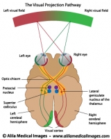
Visual pathways , labeled diagram.
Diagram of visual projection pathway from eyes to brain (visual cortex), labeled.
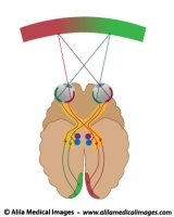
Visual pathways , unlabeled diagram.
Diagram of visual projection pathway from eyes to brain (visual cortex), unlabeled.
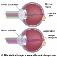
Astigmatism, labeled diagram.
The oval shape of the cornea of the eye in people with astigmatism creates multiple focal points.
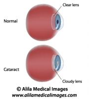
Cataract, labeled diagram.
Illustration of Eye disease - Cataract, clouding of lens.
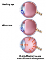
Glaucoma, unlabeled diagram.
Illustration of stages of development of glaucoma, a common eye disease.
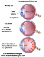
Glaucoma, labeled diagram.
Illustration of stages of development of glaucoma, a common eye disease.
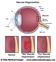
Macular degeneration, labeled diagram.
Diagram showing eye common disease: macular degeneration (dry and wet), labeled.
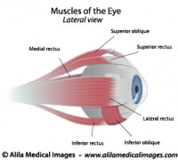
Muscles of the eye, unlabeled diagram.
Diagram showing extrinsic muscles controlling movement of the eye, unlabeled.
Muscles of the eye, labeled diagram.
Diagram showing extrinsic muscles controlling movement of the eye, labeled.
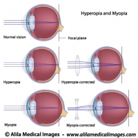
Common eye defects diagrams
Myopia, hyperopia and correction with lenses, labeled illustrations.
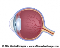
Eye anatomy unlabeled diagram.
Cross section of human eye illustration, unlabeled.
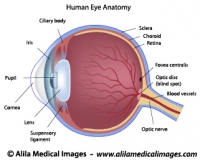
Eye anatomy labeled diagram.
Cross section of human eye illustration, labeled.
Eye adaptation to near vision, illustration.
The near response of the eye , lens accommodation, labeled diagram.
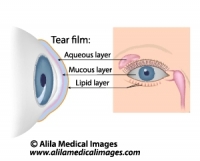
Dry eye syndrome, labeled diagram
Tears composition and the glands involved in tears production, labeled.


Email this to someoneShare on FacebookTweet about this on TwitterShare on Google+Share on LinkedIn

Ear, Hearing Gallery

Download medical drawings of human ear and cochlea anatomy, auditory pathway and mechanism of hearing, middle ear infection.

Please note: Free downloads are intended to facilitate healthcare education for people in need in low income countries and can be used for educational purposes only. If you can afford it or if you plan to use the images for commercial purposes, please consider buying instead. You can find a larger image collection at higher resolutions for sale at affordable prices on Alila Medical Media website.

To download: right click on full size image – choose “Save image as” and save it into your computer. By downloading from this website you acknowledge that you agree to our Conditions of Use.
To purchase larger sizes of the image: (left) click on full size image.

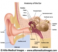
Ear anatomy, labeled diagram.
Human ear anatomy showing outer, middle and inner ear, eardrum, ossicles and cochlea, labeled drawing.
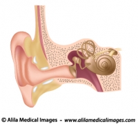
Ear anatomy, unlabeled diagram.
Human ear anatomy showing outer, middle and inner ear, eardrum, ossicles and cochlea, unlabeled drawing.
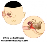
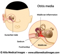
Middle ear infection, unlabeled diagram.
Otitis media diagram - infection of the middle ear, a common condition in children, unlabeled.
Middle ear infection, labeled diagram.
Otitis media diagram - infection of the middle ear, a common condition in children, labeled.
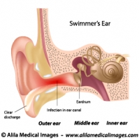
Outer ear infection, labeled diagram.
Illustration of Swimmer's ear - inflammation of outer ear canal.
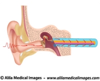
Hearing mechanism, unlabeled diagram.
Diagram showing how sound waves are transmitted and converted into vibration along the basilar membrane, the cochlea is straightened to simplify.
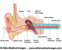
Hearing mechanism, labeled diagram.
Diagram showing how sound waves are transmitted and converted into vibration along the basilar membrane, the cochlea is straightened to simplify.


Email this to someoneShare on FacebookTweet about this on TwitterShare on Google+Share on LinkedIn

Molecular, Cell biology & Genetics Gallery

Download diagrams of normal human genome and chromosome changes in genetic diseases, illustrations of stem cells and cellular differentiation, gene expression and signaling, intracellular organelles, molecular techniques and more.

Please note: Free downloads are intended to facilitate healthcare education for people in need in low income countries and can be used for educational purposes only. If you can afford it or if you plan to use the images for commercial purposes, please consider buying instead. You can find a larger image collection at higher resolutions for sale at affordable prices on Alila Medical Media website.

To download: right click on full size image – choose “Save image as” and save it into your computer. By downloading from this website you acknowledge that you agree to our Conditions of Use.
To purchase larger sizes of the image: (left) click on full size image.

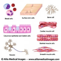
Human cell collection
Human cell collection, diagrams
DNA fingerprinting, diagram.
DNA fingerprinting, in this example 6 loci are tested, molecular weight marker and allelic ladders added.
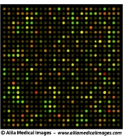
DNA microarrays chip, medical drawing.
Typical DNA chip results, technique used in (for example) cancer research to study differential expression of genes, illustration unlabeled.
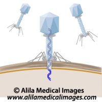
Phage infecting a bacterium, unlabeled diagram.
Phage injecting DNA into a bacterium, unlabeled drawing.
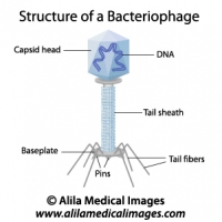
Structure of a bacteriophage, labeled diagram.
Structure of a phage, a virus that infects bacteria.
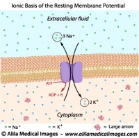
Ionic basis of resting membrane potential, labeled diagram.
Resting membrane potential is maintained by sodium potassium pump.
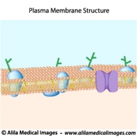
Structure of plasma membrane, unlabeled diagram.
Structure of cell membrane with lipid bilayer, cholesterol, transmembrane proteins and pores.
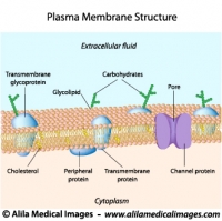
Structure of plasma membrane, labeled diagram.
Structure of cell membrane with lipid bilayer, cholesterol, transmembrane proteins and pores.
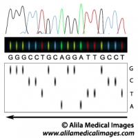
DNA sequencing principle, illustration.
DNA sequencing gel and interpretation of DNA sequence.
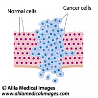
Cancer cells in a growing tumor, labeled diagram.
Cancer cells grow and break through the basement membrane, labeled drawing.
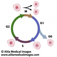
Cell cycle diagram.
Cell cycle diagram.
Phases of Cell cycle: mitosis, gap 1 (G1), gap 2 (G2), and S - DNA synthesis, G0 = quiescence.
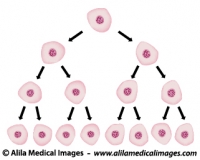
Cell division illustration.
Exponential growth of cells in a tissue, doubling of numbers of cells, unlabeled.
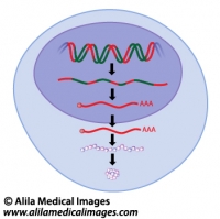
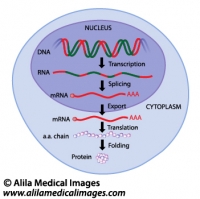
Gene expression, unlabeled diagram.
Gene expression process in eukaryotes, from transcription to protein folding.
Gene expression, labeled diagram.
Gene expression process in eukaryotes, from transcription to protein folding.
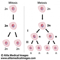
Mitosis versus meiosis, diagram.
comparing the two different types of cell division.
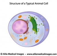
A typical cell, unlabeled diagram.
Structure of a typical eukaryotic cell, unlabeled drawing.
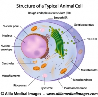
A typical cell, labeled diagram.
Structure of a typical eukaryotic cell, labeled drawing.
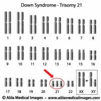
Down syndrome karyotype diagram.
Down syndrome karyotype, extra chromosome 21.
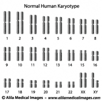
Normal human karyotype diagram.
Normal human diploid set of chromosomes.
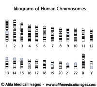
Human chromosome idiograms, diagrams
Accurate human chromosome banding patterns.
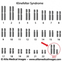
Klinefelter syndrome genome diagram.
Klinefelter syndrome genome has an extra X chromosome, XXY.
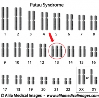
Patau syndrome, trisomy 13 diagram.
Patau syndrome genome has an extra chromosome 13.
Spermatogenesis process, labeled diagram.
Process of sperm production and meiosis in seminiferous tubule, labeled diagram.
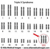
Triple X syndrome genome diagram.
Trisomy X syndrome genome has an extra X chromosome, female XXX.
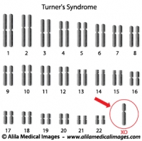
Turner's syndrome genome diagram.
Monosomy X, Turner's syndrome genotype is XO, one X chromosome is missing, female phenotype.
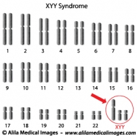
Super Male Syndrome genome diagram.
XYY, Super Male Syndrome genome has an extra Y chromosome.


Email this to someoneShare on FacebookTweet about this on TwitterShare on Google+Share on LinkedIn

Immune System Gallery

Download illustrations of cellular and humoral immunity, AIDS and HIV diagrams, cliparts of T cells and B cells in inflammation response and allergy. Also includes autoimmune diseases. The lymphatic system has its own gallery and is not included in here.

Please note: Free downloads are intended to facilitate healthcare education for people in need in low income countries and can be used for educational purposes only. If you can afford it or if you plan to use the images for commercial purposes, please consider buying instead. You can find a larger image collection at higher resolutions for sale at affordable prices on Alila Medical Media website.

To download: right click on full size image – choose “Save image as” and save it into your computer. By downloading from this website you acknowledge that you agree to our Conditions of Use.
To purchase larger sizes of the image: (left) click on full size image.

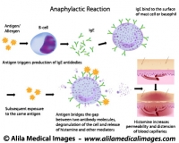
Mechanism of allergy (drug,hay fever, asthma...)
Anaphylactic reaction mechanism: antigen bridges the gap between two antibody molecules on mast cell or basophil, triggering massive release of histamine and other mediators.
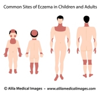
Sites of eczema on body, medical drawing.
Common sites of eczema in children and adults, medical illustration unlabeled.
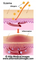
Eczema, allergic reaction, labeled diagram.
Allergens induce inflammation response of skin causing rash, redness and swelling.
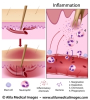
Inflammation process, labeled diagram.
Anatomy of a splinter: chemical signals dilate nearby blood vessels, neutrophils adhere to the inside of blood vessel wall (margination), squeeze out between its endothelial cells (diapedesis), chemotaxis to the site of damage and phagocytose the bacteria.
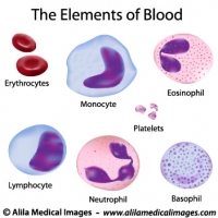
Elements of blood, medical drawing.
Illustration of red and white blood cells, lymphocytes, monocytes, platelets, basophil, eosinophil, neutrophil,.
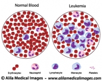
Leukemia versus normal blood count, illustration.
Cancer of blood, Anemia, medical drawing.
Antigen presenting cell functions, labeled diagram.
APC cells engulf enemy cell by phagocytosis and present pieces of antigen on their surface for recognition by T helper cells.
Function of T helper cells, labeled diagram.
Immune system basics: activation and action of T helper cells.
Cytotoxic T cells functions, labeled diagram.
Immune system basics, activation and action of cytotoxic t cells.
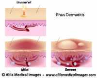
Contact dermatitis, medical drawing.
Urushiol oil induced contact (Rhus) dermatitis, illustration of the allergic reaction, unlabeled.
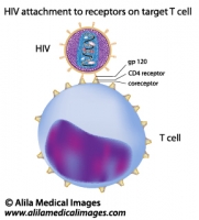
HIV attachment to target T cell, labeled diagram.
HIV attaches to target T cell through gp120 and CD4 receptor and coreceptor on host cell.
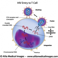
HIV entry to T cell, labeled drawing.
HIV attachment and entry to T cell and becomes a provirus. Labeled diagram


Email this to someoneShare on FacebookTweet about this on TwitterShare on Google+Share on LinkedIn