Tag Archives: endocrinology

The Hypothalamus and Pituitary Gland, with Animation.

This video is available for licensing on our website. Click HERE!

The hypothalamus and the pituitary gland are at the center of endocrine function. The hypothalamus is part of the brain, while the pituitary, also called hypophysis (hy-POFF-ih-sis), is an endocrine gland. The hypothalamus links the nervous system to the endocrine system via the pituitary gland. The two structures are located at the base of the brain and are connected by a thin stalk.

The hypothalamus produces several hormones, known as neurohormones, which control the secretion of other hormones by the pituitary. Pituitary hormones, in turn, control the production of yet other hormones by other endocrine glands.

The pituitary has two distinct lobes:

The anterior pituitary, also called adenohypophysis (AD-eh-no-hy-POFF-ih-sis), communicates with the hypothalamus via a network of blood vessels known as the hypophyseal portal system. Several neurohormones produced by the hypothalamus are secreted into the portal system to reach the anterior pituitary, where they stimulate or inhibit production of pituitary hormones. Major hormones include:

  • Gonadotropin-releasing hormone, GnRH, a hypothalamic hormone, stimulates the anterior pituitary to producefollicle-stimulating hormone, FSH, and luteinizing hormone, FSH and LH, in turn, control the activities of the gonads – the ovaries and testes.
  • Corticotropin-releasing hormone, CRH,promotes the secretion of adrenocorticotropic hormone, ACTH, which in turn stimulates production of cortisol by the adrenal gland.
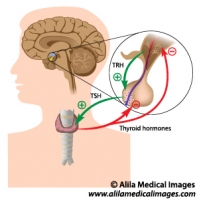
  • Thyrotropin-releasing hormone, TRH, promotes the release of thyroid-stimulating hormone, TSH, and prolactin. TSH, in turn, induces the thyroid gland to produce thyroid hormones. Prolactin stimulates the mammary glands to produce milk.
  • Prolactin-inhibiting hormone, PIH, inhibits production of prolactin.
  • Growth hormone–releasing hormone, GHRH, promotes production of growth hormone, or somatotropin, which has widespread effects on the growth of various tissues in the body.
  • Growth hormone–inhibiting hormone, GHIH, or somatostatin, inhibits production of growth hormone.

The posterior pituitary, also called neurohypophysis, communicates with the hypothalamus via a bundle of nerve fibers. These are essentially hypothalamic neurons with cell bodies located in the hypothalamus while their axons EXTENDED to posterior pituitary. These neurons produce hormones, transport them down the stalk, and store them at the nerve terminals within the posterior pituitary, where they wait for a nerve signal to trigger their release. Two hormones have been identified so far:

– Vasopressin, also known as antidiuretic hormone, ADH, acts on the kidneys to retain water.

– Oxytocin causes the uterus to contract during childbirth and stimulates contractions of the milk ducts in lactating women.

Email this to someoneShare on FacebookTweet about this on TwitterShare on Google+Share on LinkedIn

Endocrine System Gallery

Download diagrams illustrating anatomy of endocrine organs, hypothalamus and pituitary gland, steroid and non-steroid hormones actions, signaling pathways, endocrine disorders.

Please note: Free downloads are intended to facilitate healthcare education for people in need in low income countries and can be used for educational purposes only. If you can afford it or if you plan to use the images for commercial purposes, please consider buying instead. You can find a larger image collection at higher resolutions for sale at affordable prices on Alila Medical Media website.

To download: right click on full size image – choose “Save image as” and save it into your computer. By downloading from this website you acknowledge that you agree to our Conditions of Use.
To purchase larger sizes of the image: (left) click on full size image.

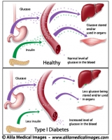
Type 1 diabetes, labeled diagram.
Type 1 diabetes, insulin dependent: pancreas does not produce enough insulin, medical drawing.
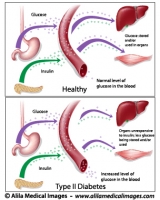
Type 2 diabetes, labeled diagram.
Type 2 diabetes, insulin resistance, pathology of the disease, labeled drawing.
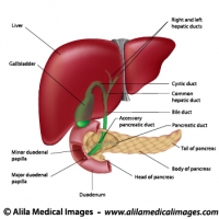
Digestive organs and bile ducts, labeled diagram.
Diagram of human liver, gallbladder, duodenum and pancreas anatomy, labeled.
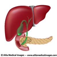
Digestive organs, medical drawing.
Diagram showing anatomy of liver pancreas gallbladder and bile ducts, unlabeled version.
Diabetes type 1, labeled diagram.
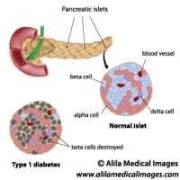
Anatomy of the pancreas with details of pancreatic islet: normal versus type 1 diabetic where beta cells are destroyed.
Polycystic and normal ovary, labeled diagram.
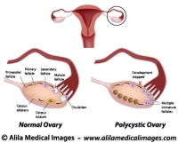
Normal ovarian cycle and Polycystic ovary syndrome illustration, labeled.
Adrenal gland, unlabeled drawing.
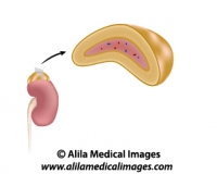
The adrenal gland location and anatomy, unlabeled diagram
Adrenal gland, labeled drawing.
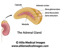
The adrenal gland location and anatomy, labeled diagram
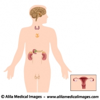
The endocrine system, unlabeled diagram.
Anatomy of endocrine organs in male and female, unlabeled diagram.
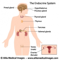
The endocrine system, labeled diagram.
Anatomy of endocrine organs in male and female, labeled diagram.
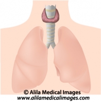
Thyroid gland, medical drawing.
Thyroid gland location and anatomy, unlabeled.
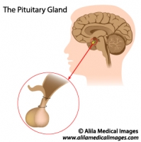
Pituitary gland, medical drawing.
Illustration of the pituitary gland, anterior and posterior lobes, unlabeled version.
The pituitary thyroid axis, labeled diagram.
Negative feedback in the pituitary thyroid axis: thyroid hormones repress their own secretion.
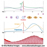
Menstrual cycle and ovulation, labeled diagram.
Normal menstrual cycle and the hormonal control of ovulation, labeled illustration.


Email this to someoneShare on FacebookTweet about this on TwitterShare on Google+Share on LinkedIn