Tag Archives: hair problems

Common ankle injuries

Ankle anatomy

The ankle includes three bones : the tibia (shinbone), the fibula and the talus (Fig. 1). Articulations between these bones make up the ankle joint.

Ankle joint labeled diagram.
Fig.1: Anatomy of the ankle joint.  Click on image to see a larger version on  Alila Medical Media website where the image is also available for licensing.


The ankle is stabilized by the following ligaments (Fig. 2):
– tibiofibular ligaments connect the tibia to the fibula, one in front (anterior tibiofibular) and one in the back (posterior tibiofibular);
– lateral collateral ligaments connect the fibula to the talus (two of them: again one in front and one in the back) and to the calcaneus (the heel bone); and
– on the medial side, a multipart deltoid ligament connects the tibia to the talus and other bones of foot (the calcaneus and navicular).

Ligaments of ankle labeled.
Fig.2 : Ligaments of ankle.  Click on image to see a larger version on  Alila Medical Media website where the image is also available for licensing.


Common ankle injuries include ankle sprains and ankle fractures.

Ankle sprain

Ankle sprain refers to injury to any of the ligaments of the ankle joint. This happens when the  ankle is rolled or twisted beyond the normal range of motion and the ligaments are overstretched and torn. Commonly, the ankle moves suddenly outward while the foot turns inward resulting in overstretching of the ligaments on the outside of the foot (lateral ligaments). This type of sprain is called inversion (Fig. 3). On the other hand, when the ankle moves inwards and the foot turns outwards it’s the ligaments on the inside (medial) that are hurt. This type of sprain is called eversion and is much less common.
Ankle sprains
Fig.3 :Types of ankle sprain, illustrated for the right foot, anterior view. Click on image to see a larger version on Alila Medical Media website where the image is also available for licensing.


Ankle sprains are common sport injuries. They can range from mild to severe depending on how bad is the damage and how many ligaments are involved (Fig. 4)

Ankle sprain grading
Fig.4 :Grades of ankle sprain. Click on image to see a larger version on Alila Medical Media website where the image is also available for licensing.


In most cases sprains can be treated with rest, ice pack, compression with a bandage and elevation (raising your foot up) to reduce swelling. Severe injuries may require surgery.

Ankle fracture

Broken bones of the ankle, a common sport injury. Commonly due to a direct blow to the ankle or a fall. Pott’s fracture (Fig. 5) represents a typical situation when the ankle receives a blow from the outside resulting in broken fibula at the point of impact. The talus moves outward shearing off a piece of the tibia. Medial ligaments are also injured in this case.

Pott's fracture labeled.
                                                                                   Fig. 5: Pott’s fracture. Click on image to see a larger version on Alila Medical Media website where the image is also available for licensing.


Imaging techniques such as x-ray or CT-scan are used to determine the severity of fractures. If the broken bones are still in their normal position they will be immobilized (with a cast for example) to facilitate healing. Bones that are fallen out of place will require surgery. During surgery the bones are positioned back to their normal place, screws and metal plates are then used to keep the fragments together.

                                                                                                              >  See all Orthopedic topics

Email this to someoneShare on FacebookTweet about this on TwitterShare on Google+Share on LinkedIn

Heart, Blood and Circulation Gallery

Download illustrations of human circulatory system: gross anatomy, cardiac cycle, heart diseases and congenital defects, cardiovascular conditions and surgery treatment, blood cell formation, vascular disorders, and more.

Please note: Free downloads are intended to facilitate healthcare education for people in need in low income countries and can be used for educational purposes only. If you can afford it or if you plan to use the images for commercial purposes, please consider buying instead. You can find a larger image collection at higher resolutions for sale at affordable prices on Alila Medical Media website.

To download: right click on full size image – choose “Save image as” and save it into your computer. By downloading from this website you acknowledge that you agree to our Conditions of Use.
To purchase larger sizes of the image: (left) click on full size image.

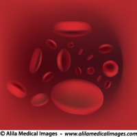
Blood flow in vessel, medical drawing.
Red blood cells streaming in artery
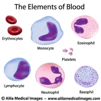
Elements of blood, medical drawing.
Illustration of red and white blood cells, lymphocytes, monocytes, platelets, basophil, eosinophil, neutrophil.
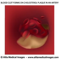
Atherosclerosis, unlabeled.
Blood clot forms on cholesterol plaque in artery blocking blood flow.
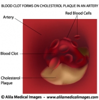
Atherosclerosis, labeled.
Blood clot forms on cholesterol plaque, blocked artery.
Process of cholesterol plaque in artery, unlabeled
Diagram showing process of cholesterol plaque in artery, unlabeled version.
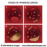
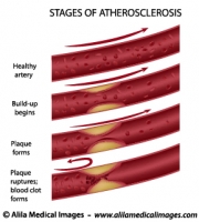
Stages of Atherosclerosis
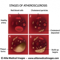
Process of cholesterol plaque build-up in artery, labeled diagram.
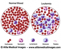
Leukemia versus normal blood count, illustration.
Cancer of blood, Anemia, medical drawing.
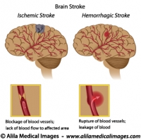
Brain stroke diagrams
Ischemic versus Hemorrhagic brain stroke, unlabeled.
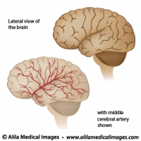
Blood supply to the brain diagram
Lateral view of the brain with middle cerebral artery, unlabeled.
Stages of atherosclerosis diagram
Cholesterol plaque build-up in blood vessel and formation of blood clot cause blockage of blood flow, labeled.
Clipart of healthcare icon (cardiology)
Magnifying glass on an ECG graph diagram
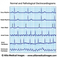
Cardiac arrhythmias ECG/ EKG set diagram.
Normal and pathological electrocardiograms in heart diseases.
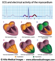
EKG and myocardium function diagram
Phases of electrical activity of human heart, ECG cycle.
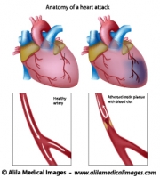
Myocardial infarction diagram (heart attack)
Coronary artery blocked by cholesterol plaque causing heart stroke.
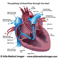
Human heart blood flow
Illustration of blood flow through the heart, labeled
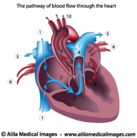
Human heart blood flow
Illustration of blood flow through the heart, unlabeled
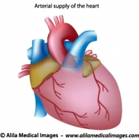
Blood supply to the heart unlabeled diagram
Coronary arteries illustration, anterior view of the heart
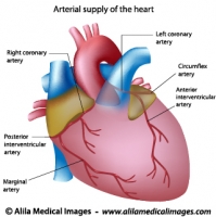
Blood supply to the heart labeled diagram
Coronary arteries illustration, anterior view of the heart
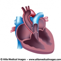
Human heart cut, medical illustration.
Heart anatomy diagram, unlabeled.
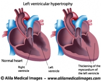
Left ventricular hypertrophy labeled diagram
Thickening of the myocardium of left ventricle of human heart.
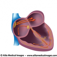
Electrical pathways of the heart, unlabeled
The cardiac conduction system of human heart, unlabeled version.
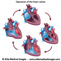
Heart valves operation, unlabeled diagram.
Cycles of heart valves operation pumping blood through different chambers, aorta and veins.
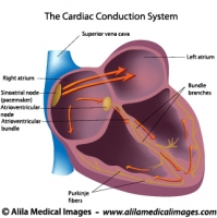
Electrical pathways of the heart
The cardiac conduction system of human heart, labeled version.
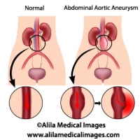
Abdominal aortic aneurysm diagram
Aneurysm in aorta of the abdomen and bleeding risk.
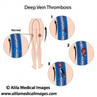
Deep vein thrombosis diagram
Abnormal blood clotting in deep leg vein and formation of thrombus.
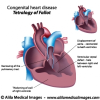
Tetralogy of Fallot diagram
Congenital heart disease: Tetralogy of Fallot, labeled
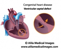
Ventricular septal heart defect diagram.
Congenital heart disease: ventricular septal defect, hole in the septum between ventricles, unlabeled.
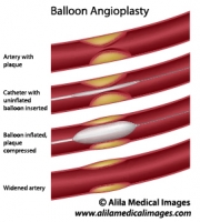
Balloon angioplasty procedure diagram
Heart cath procedure with balloon to widen artery with plaque
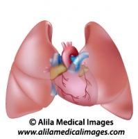
Heart and lungs diagram unlabeled
Human lungs and heart anatomy, unlabeled.
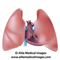
Thorax anatomy unlabeled diagram.
Human heart cross section and lungs anatomy.
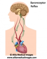
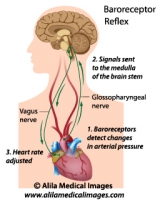
Biofeedback regulation of heart rate, medical illustration.
Regulation of blood pressure through baroreflex, unlabeled version.
Biofeedback regulation of heart rate, labeled.
Regulation of blood pressure through baroreflex, labeled version.
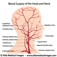
Blood supply to the head and neck, labeled drawing.
Diagram of human head anatomy with arteries including internal carotid supplying the brain, lateral (side) view, labeled.
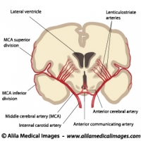
Blood supply to the brain, coronal section.
Middle cerebral artery anatomy and distribution, labeled diagram.
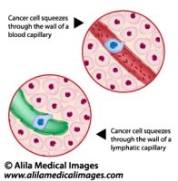
Metastatic cancer, medical drawing.
Cancer cell squeezes through the endothelium of blood and lymph vessel, illustration unlabeled.
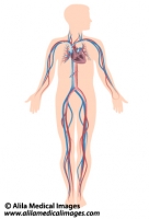
Human circulatory system diagram
Anatomy of human circulation system, unlabeled.
Email this to someoneShare on FacebookTweet about this on TwitterShare on Google+Share on LinkedIn

Skin and Hair Gallery

Download diagrams of human epidermis, dermis and hair follicle anatomy, skin problems and diseases, plastic surgery procedures.

Please note: Free downloads are intended to facilitate healthcare education for people in need in low income countries and can be used for educational purposes only. If you can afford it or if you plan to use the images for commercial purposes, please consider buying instead.  You can find a larger image collection at higher resolutions for sale at affordable prices on Alila Medical Media website.

To download: right click on full size image – choose “Save image as” and save it into your computer. By downloading from this website you acknowledge that you agree to our Conditions of Use.
To purchase larger sizes of the image: (left) click on full size image.

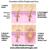
Skin acne formation, labeled diagram.
Anatomy of human skin showing process of formation of skin acne and pimples.
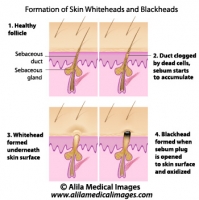
Skin blackheads illustration
Formation of skin blackhead and whitehead, labeled diagram.
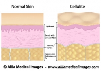
Cellulite versus smooth skin, labeled diagram.
Illustration of normal skin anatomy (epidermis, dermis and subcutaneous fat) and changes in skin with cellulite : accumulation of fat cells push the fibrous septae up resulting in skin dimpling - bumps. Also, collagen fibers are less in number and less organised.
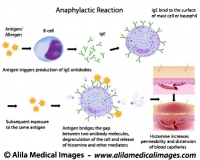
Mechanism of allergy (drug,hay fever, asthma...)
Anaphylactic reaction mechanism: antigen bridges the gap between two antibody molecules on mast cell or basophil, triggering massive release of histamine and other mediators.
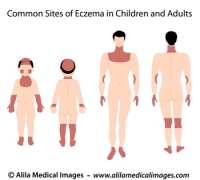
Sites of eczema on body, medical drawing.
Common sites of eczema in children and adults, medical illustration unlabeled.
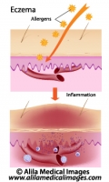
Eczema, allergic reaction, labeled diagram.
Allergens induce inflammation response of skin causing rash, redness and swelling.
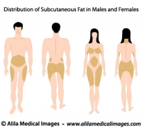
Subcutaneous fat distribution diagram.
Subcutaneous fat distribution in men and women, medical illustration.
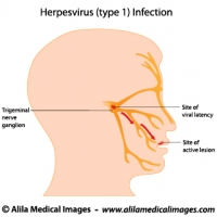
Herpes simplex virus infection, labeled diagram.
Herpes simplex virus (HSV-1) latent (in trigeminal nerve ganglion) and active infection (cold sores), labeled drawing.
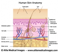
Human skin anatomy, labeled.
Structure of human skin, epidermis, dermis and hypodermis layers with hair follicle, sebaceous glands, nerve endings and blood supply.
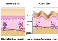
Skin aging, medical illustration.
Older skin with wrinkles, age spots, dermis and hypodermis atrophy, reduced and disorganized collagen fibers.
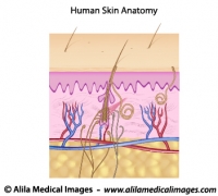
Human skin anatomy, unlabeled.
Structure of human skin, epidermis, dermis and hypodermis layers with hair follicle, sebaceous glands, nerve endings and blood supply.
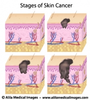
Skin cancer staging, medical drawing.
Melanoma of skin, staging based on layers affected (epidermis, dermis, hypodermis) and presence of metastasis, unlabeled.
Stages of pressure sores, labeled diagram.
Anatomy of skin layers and soft tissue showing different degrees of bed sores. Labeled illustration.
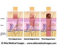
Degrees of skin burns, medical illustration.
First degree of burn affect mostly the epidermis, second degree affects the dermis and third goes deeper into the hypodermis and below.
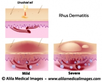
Contact dermatitis, medical drawing.
Urushiol oil induced contact (Rhus) dermatitis, illustration of the allergic reaction, unlabeled.


Email this to someoneShare on FacebookTweet about this on TwitterShare on Google+Share on LinkedIn