Tag Archives: respiratory system

Paranasal sinuses and sinus conditions

The videos on this page can be downloaded upon purchase of a license on Alila Medical Media website. Click here!


Paranasal sinuses, or simply “sinuses” in common language,  are air cavities in the bones of the skull. There are four pairs of sinuses (see Fig. 1, 2 and upper panel of Fig. 3):

– the maxillary sinuses are under the eyes, in the maxillary bones.
– the frontal sinuses are above the eyes, in the frontal bone.
– the ethmoid sinuses are between the nose and the eyes, in the ethmoid bone.
– the sphenoid sinuses are behind the nasal cavity, in the sphenoid bones.

Sinusitis
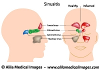
Fig.1: The four pairs of sinuses. Red = frontal, green =  ethmoid, blue = sphenoid, beige =  maxillary. The right panel show normal sinuses on half of the head and inflamed sinuses on the other half. Click on image to see a larger version on Alila Medical Media website where the image is also available for licensing.


The sinuses are lined with respiratory epithelium producing mucus. The mucus drains into nasal cavity through small openings (Fig. 2 left panel, Fig. 3 upper panel). Impaired sinus drainage has been associated with inflammation of sinuses (sinusitis, see below).
Biological function of the sinuses remains unclear.

Nose anatomy labeled. .
Fig. 2: Front view of the sinuses (left panel) showing connections to the nasal cavity. Right panel shows mid-sagittal section of the head. Click on image to see a larger version on Alila Medical Media website where the image is also available for licensing.


Sinusitis or rhinosinusitis is inflammation of the paranasal sinuses (Fig. 1, right panel). This can be due to:
– allergy (allergic rhinitis): allergens such as pollen, pet dander,.. trigger overreaction of the mucosa of the nose and sinuses resulting in excess mucus, nasal congestion, sneezing and itching.
– infection: usually as a complication of an earlier viral infection of the nasal mucosa, pharynx or tonsils such as during a common cold. Impaired sinus drainage due to inflammation of nasal mucosa during a cold often leads to infection of the sinus itself. Cold-like symptoms plus headache and facial pain/pressure are common complaints.
– other conditions that cause blockage of sinus drainage: structural abnormality such as deviated nasal septum (Fig. 3); formation of nasal polyps (Fig. 4). When a sinus is blocked, fluid builds up making it a favorable environment for bacteria, viruses or fungi to grow and cause infection.
Deviated nasal septum
Fig. 3: Front view of the sinuses (upper panel) showing connections to the nasal cavity, also shown the nasal septum (light blue color). Lower panel shows deviated septum blocking drainage of the right maxillary sinus (your left). Click on image to see a larger version on Alila Medical Media website where the image is also available for licensing.


Nasal polyps

Fig. 4: Nasal polyps – overgrowths of nasal mucosa – block sinus drainage. Click on image to see a larger version on Alila Medical Media website where the image is also available for licensing.


Treatment depends on the cause of sinusitis:
– For viral infection : symptom relief medications such as nasal spray for irrigation and decongestion; other conservative treatment for common cold such as rest and drinking plenty of fluid.
– For bacterial infection: antibiotics may be prescribed.
– For allergy: intranasal corticosteroids are commonly used.
– For recurrent (chronic) sinusitis due to structural abnormalities or nasal polyps, nasal surgery may be recommended.

                                                                                                           See all Respiratory topics

Email this to someoneShare on FacebookTweet about this on TwitterShare on Google+Share on LinkedIn

Asthma (with Animation Video)

The videos on this page can be downloaded upon purchase of a license on Alila Medical Media website. Click here!


Asthma is a chronic respiratory condition where the airway is inflamed and narrowed causing breathlessness, wheezing, chest tightness and coughing. Symptoms come as recurrent episodes called asthmatic attacks more commonly during the night and early morning. Asthma is usually diagnosed in childhood and lasts for life.

Anatomy of asthma

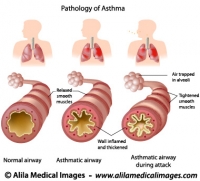
Our lungs consist of millions of air tubes or airways (bronchi and smaller bronchioles) bringing air in and out of the body. Each tube ends with a cluster of air sacs (alveolus, plural alveoli) where the gas exchange process takes place. The airways have a layer of smooth muscle in their wall which enables them to constrict or dilate. In response to the body’s higher demand of air, such as during exercise, the airways dilate to increase air flow. In response to presence of pollutants in the air, they constrict to prevent the lungs from being polluted. In people suffering from asthma these airways are inflamed, narrowed and become more sensitive to certain substances. Asthmatic attack (or exacerbation) happens when the airways react to these substances. During the attack smooth muscle contracts squeezing the airways making them even narrower, mucus secretion increases further obstructs the airways.

Causes of asthma

Causes of asthma are complex and not fully understood but likely involve a combination of genetic and environmental factors. Family history is a known risk factor for asthma. There are at least over twenty genes associated with asthma of which many are involved in the immune system. Most people who have asthma also have allergies. Many environmental factors such as air pollution, chemicals, smoking, allergens have been associated with development of asthma or triggering of asthmatic attacks.

Triggers of asthmatic attack

Triggers are factors that initiate the attack, these can be very different from person to person. Common triggers include :
– allergens (pollen, animal fur, pet dander, sulfites in preserved food..)
– irritants (cigarette smoke, industrial chemicals, dust, household chemicals,..)
– some medication (aspirin, beta blockers,..).
– physical activity, exercise.

Treatments

There is no cure for asthma. The most effective way to manage symptoms is to identify the triggers of asthmatic attack and avoid them.
There are two main classes of medication:
– bronchodilators – substances that dilate bronchi and bronchioles – are used as short-term relief of symptoms.
– inflammation moderators such as corticosteroids are used as long-term treatment.
Asthma inhalers are used to deliver medication to the lungs.

Associated conditions

A number of conditions tend to occur more frequently in people with asthma:
– Allergies :  eczema and hay fever. These individuals are considered hyperallergic (high tendency to develop allergic reactions). The combination of these conditions is called atopy or atopic symdrome.
Gastroesophageal reflux disease (GERD): a condition in which stomach acid backs up and damages the mucosal lining of the esophagus. GERD may worsen asthma symptoms and medications for asthma often worsen GERD symptoms. Treating GERD usually improves asthma and must be included in asthma treatment plan.
Obstructive sleep apnea (OSA): asthmatic patients tend to develop OSA. The mechanism is not fully understood but it’s likely due to nasal obstruction. Click on the link to read more about OSA.
Sinusitis: inflammation of paranasal sinuses. Sinusitis commonly worsens asthma symptoms and makes treatment less effective.

                                                                                                        See all Respiratory topics

                                                                                                        See all Immunology topics


Email this to someoneShare on FacebookTweet about this on TwitterShare on Google+Share on LinkedIn

Snoring and Sleep Apnea

Below is a narrated  animation of snoring, sleep apnea and treatment with a mandibular advancement device. Click here to license this video and/or other related videos on Alila Medical Media website.


No one likes to share a bedroom with a person who snores. Snoring is definitely well established as a social/marital problem. Not many are aware, however, that snoring maybe a sign of, or might progress to, a more serious, potentially life threatening health problem: obstructive sleep apnea (OSA).
Sleep apnea is a sleep disorder characterized by instances of cessation of breathing (apnea).

Anatomy of snoring and sleep apnea

In normal breathing, air enters the nostrils and goes through the throat and the trachea (the windpipe) to the lungs (blue arrow in Fig. 1). In people who snore this airway is partially obstructed by excess tissue of the throat (large tonsils, soft palate, tongue). Another common cause of obstruction is the dropping of the tongue into the throat due to over- relaxation of tongue muscles during sleep. To get enough air the body reacts by breathing through the mouth. The two air currents from the nose and the mouth competing through narrow spaces in the throat cause the soft palate (essentially a piece of soft tissue hanging in the throat) to vibrate. This vibration is the source of the noise we hear when someone is snoring.


Snoring and sleep apnea

Fig. 1: Anatomy of snoring and sleep apnea. Click on image to see a larger version on  Alila Medical Media website where the image is also available for licensing.


Sleep apnea happens when the airway is completely obstructed, no air can go through and the person stops breathing (apnea). This cessation of breathing triggers the brain to response by waking up the person just enough to take a breath. This repeats itself again and again during the course of the night and may result in sleep deprivation.

How do I know if i have sleep apnea?

Not everyone who snores has sleep apnea. It’s hard to self diagnose when one is sleeping. The biggest tell-tale to look for is daytime drowsiness together with other signs of sleep deprivation such as loss of concentration, loss of memory, headache in the morning,…It would also help to have a bed partner observing for episodes of breathing pauses.  If you suspect that you may have sleep apnea  based on those symptoms, it’s important that you visit your doctor immediately. The doctor will examine your throat and if a sleep disorder is suspected, you will be sent to a sleep clinic where your sleep will be monitored and data about your breathing patterns will be collected.

Treatments for snoring and sleep apnea

(in order from mild to severe)

Life style changes: Snoring and mild sleep apnea may be treated with life style changes such as losing some weight, avoiding alcohol and heavy meals. In some people, sleeping on the side instead of on the back might be a solution.
Oral appliances (snoring mouthpiece) are small devices that can be worn in the mouth at night. An oral appliance prevents the tongue and other soft tissue from falling back into your throat and thus keeps the airway open. It’s best to have the device made to measure to ensure fitting to your mouth.
Continuous Positive Airway Pressure (CPAP) is the first line treatment for patients with severe OSA. In this case a small machine is used to generate a constant air flow that is delivered to the patient through a mask fitted on the nose. It looks pretty much like a snorkeling mask except that the tube is connected to a machine.
It is important to note that CPAP and oral appliances help to keep your airway open and enable you to sleep at night but they do not cure the condition. The symptoms will return as soon as you stop using them.
Surgery: Various surgical procedures are available for treatment of sleep apnea. Surgery can be performed on the soft palate, the tongue, the hyoid bone, the jaws or a combination of those. Your surgeon will advice which is best for each patient.

                                                                                                                           See all ENT topics

Email this to someoneShare on FacebookTweet about this on TwitterShare on Google+Share on LinkedIn

Respiratory System Gallery

Download anatomical drawings of upper and lower human respiratory tract, diagrams of breathing mechanism and pulmonary function, lung diseases, conditions, problems caused by smoking. Also includes sinuses, larynx (voice box) anatomy and disorders.

Please note: Free downloads are intended to facilitate healthcare education for people in need in low income countries and can be used for educational purposes only. If you can afford it or if you plan to use the images for commercial purposes, please consider buying instead. You can find a larger image collection at higher resolutions for sale at affordable prices on Alila Medical Media website.

To download: right click on full size image – choose “Save image as” and save it into your computer. By downloading from this website you acknowledge that you agree to our Conditions of Use.
To purchase larger sizes of the image: (left) click on full size image.

Anatomy of Asthma, labeled.
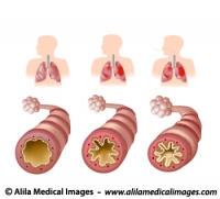
Pathology of asthma, chronic inflammatory disease of the airways, and asthmatic attack, labeled diagram.
Mechanism of allergy (drug,hay fever, asthma...)
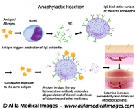
Anaphylactic reaction mechanism: antigen bridges the gap between two antibody molecules on mast cell or basophil, triggering massive release of histamine and other mediators.
Common respiratory diseases, labeled diagram.
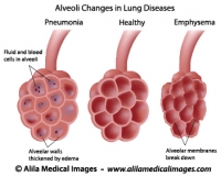
Alveoli changes in lung diseases: pneumonia and emphysema.
Common respiratory diseases, unlabeled diagram.
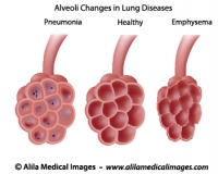
Alveoli changes in lung diseases: pneumonia and emphysema.
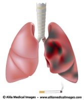
Smoker's lung (with cancer) versus healthy lung
Smoker's lung with tumor and tar deposit versus healthy lung, medical illustration.
Anatomy of Asthma, labeled.
Pathology of asthma, chronic inflammatory disease of the airways, and asthmatic attack, unlabeled diagram.
Healthcare icon (lung), medical drawing.
Magnifying glass on lung picture, lungs check-up icon.
Sinusitis, labeled diagram.
Anatomy of sinuses of the head, anterior and lateral view, showing inflammation of sinuses.
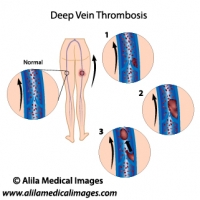
Deep vein thrombosis diagram
Abnormal blood clotting in deep leg vein and formation of thrombus.
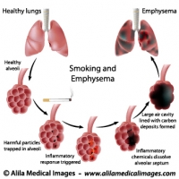
Smoking and Emphysema, labeled diagram.
Smoking tobacco damages and destroys lung alveoli, causing emphysema.
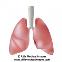
Human lungs isolated on white, drawing.
Human lungs anatomy diagram, unlabeled..
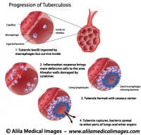
TB bacteria infection, labeled diagram.
Progression of pulmonary tuberculosis, formation of Ghon complex and tubercles.
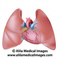
Heart and lungs diagram unlabeled
Human lungs and heart anatomy, unlabeled.
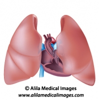
Thorax anatomy unlabeled diagram.
Human heart cross section and lungs anatomy.
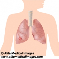
Lung cancer, medical drawing.
Tumor in lungs diagram, unlabeled.
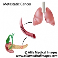
Metastatic cancer diagram
Cancer spreads through blood circulation from pancreas to lungs, unlabeled.
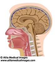
Head anatomy median section, medical drawing.
Median section of human head diagram with details of brain, sinuses, tonsils, oral and nasal cavity, pharynx and larynx, unlabeled.
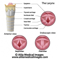
Vocal fold, labeled diagram.
Anatomy picture of the larynx, anterior and endoscopic view, showing respiration and phonation positions.
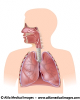
Respiratory system, unlabeled diagram
Human respiratory system diagram complete.
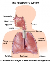
Respiratory system, labeled diagram
Human respiratory system diagram complete with labels.


Email this to someoneShare on FacebookTweet about this on TwitterShare on Google+Share on LinkedIn