Tag Archives: skin

Cellulite

The following video is available for licensing on Alila Medical Media website. Click here!


What is cellulite?

Cellulite is the dimpling, lumpy appearance of the skin, commonly occurs in females after puberty age. It’s most visible on the thighs, the buttocks, and belly. Other names include  adiposis edematosa, dermopanniculosis deformans, status protrusus cutis, gynoid lipodystrophy, orange peel syndrome and cottage cheese skin. Cellulite is not a disease and should NOT be confused with cellulitis, which is the infection of skin and underlying tissues.

Anatomy of cellulite

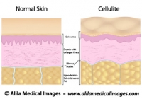
The skin has three layers : epidermis (outermost), dermis and subcutaneous fat (Fig.1). Vertical bands of connective tissue called fibrous septae (singular: septum) connect the dermis to underlying soft tissues. Cellulite happens when fat cells accumulated in the subcutaneous fat layer push the skin up while the fibrous septae pull it down. These two actions in opposite directions result in the bumpy appearance of the skin. In people with thin skin, this becomes even more noticeable.
Cellulite versus smooth skin
Fig. 1 : Structure of normal skin and skin with cellulite, back to back for comparison. Note the fibrous septae pull the skin down in cellulite. Click on image to see a larger version on Alila Medical Media website where the image is also available for licensing.


Causes

Causes of cellulite are not fully understood but the following factors are likely to be involved:
Hormonal : Over 80% of women over the age of 20 has some degree of cellulite. Cellulite is rare in men, but is more common in those with androgen deficiency.
Genetic: Some genetic make-ups are likely to be predisposing factors. You have more chance of getting it if other women in the family have it.
Lifestyle: Diet and exercises definitely have a good share of contribution. Reducing body fat typically improves cellulite appearance.  Extreme diet, however, may produce adverse effect as thinner skin makes it more visible (see the anatomy part above).

Treatment

Various therapies are available including massages, heat therapy, ultrasound, drugs,… These treatments supposedly act to either reduce subcutaneous fat or thicken the skin, but none are scientifically proven to be effective in the long term.

The latest technology based on releasing of the fibrous septae that pull the skin down (see the anatomy section above) has received a better response from scientists. Cellulaze, a device that uses laser beams to cut through the fibrous septae, has produced promising initial results in U.S. clinical trials. It’s been advised, however, to take this approach with precaution given the newness of the technique and shortness of long term data.

Finally, as repetitive and obvious as it may sound, the best treatment for cellulite is to maintain a healthy lifestyle, eating healthy (but no extreme diet), drinking lots of fluid and daily exercises.

                                                                                                  >See all dermatology topics

Email this to someoneShare on FacebookTweet about this on TwitterShare on Google+Share on LinkedIn

Skin and Hair Gallery

Download diagrams of human epidermis, dermis and hair follicle anatomy, skin problems and diseases, plastic surgery procedures.

Please note: Free downloads are intended to facilitate healthcare education for people in need in low income countries and can be used for educational purposes only. If you can afford it or if you plan to use the images for commercial purposes, please consider buying instead.  You can find a larger image collection at higher resolutions for sale at affordable prices on Alila Medical Media website.

To download: right click on full size image – choose “Save image as” and save it into your computer. By downloading from this website you acknowledge that you agree to our Conditions of Use.
To purchase larger sizes of the image: (left) click on full size image.

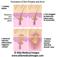
Skin acne formation, labeled diagram.
Anatomy of human skin showing process of formation of skin acne and pimples.
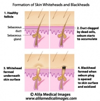
Skin blackheads illustration
Formation of skin blackhead and whitehead, labeled diagram.
Cellulite versus smooth skin, labeled diagram.
Illustration of normal skin anatomy (epidermis, dermis and subcutaneous fat) and changes in skin with cellulite : accumulation of fat cells push the fibrous septae up resulting in skin dimpling - bumps. Also, collagen fibers are less in number and less organised.
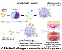
Mechanism of allergy (drug,hay fever, asthma...)
Anaphylactic reaction mechanism: antigen bridges the gap between two antibody molecules on mast cell or basophil, triggering massive release of histamine and other mediators.
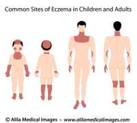
Sites of eczema on body, medical drawing.
Common sites of eczema in children and adults, medical illustration unlabeled.
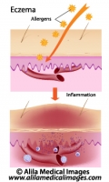
Eczema, allergic reaction, labeled diagram.
Allergens induce inflammation response of skin causing rash, redness and swelling.
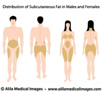
Subcutaneous fat distribution diagram.
Subcutaneous fat distribution in men and women, medical illustration.
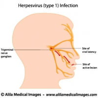
Herpes simplex virus infection, labeled diagram.
Herpes simplex virus (HSV-1) latent (in trigeminal nerve ganglion) and active infection (cold sores), labeled drawing.
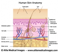
Human skin anatomy, labeled.
Structure of human skin, epidermis, dermis and hypodermis layers with hair follicle, sebaceous glands, nerve endings and blood supply.
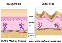
Skin aging, medical illustration.
Older skin with wrinkles, age spots, dermis and hypodermis atrophy, reduced and disorganized collagen fibers.
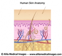
Human skin anatomy, unlabeled.
Structure of human skin, epidermis, dermis and hypodermis layers with hair follicle, sebaceous glands, nerve endings and blood supply.
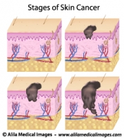
Skin cancer staging, medical drawing.
Melanoma of skin, staging based on layers affected (epidermis, dermis, hypodermis) and presence of metastasis, unlabeled.
Stages of pressure sores, labeled diagram.
Anatomy of skin layers and soft tissue showing different degrees of bed sores. Labeled illustration.
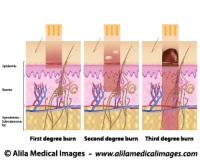
Degrees of skin burns, medical illustration.
First degree of burn affect mostly the epidermis, second degree affects the dermis and third goes deeper into the hypodermis and below.
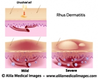
Contact dermatitis, medical drawing.
Urushiol oil induced contact (Rhus) dermatitis, illustration of the allergic reaction, unlabeled.


Email this to someoneShare on FacebookTweet about this on TwitterShare on Google+Share on LinkedIn